Explore
 Validate
 Learn
Learn Western blot
Antibody data
- Antibody Data
- Antigen structure
- References [1]
- Comments [0]
- Validations
- Western blot [1]
- Immunocytochemistry [3]
- Immunohistochemistry [3]
- Other assay [2]
Submit
Validation data
Reference
Comment
Report error
- Product number
- PA5-104528 - Provider product page

- Provider
- Invitrogen Antibodies
- Product name
- CHOP Polyclonal Antibody
- Antibody type
- Polyclonal
- Antigen
- Synthetic peptide
- Description
- Antibody detects endogenous levels of total DDIT3.
- Reactivity
- Human, Mouse, Rat
- Host
- Rabbit
- Isotype
- IgG
- Vial size
- 100 μL
- Concentration
- 1 mg/mL
- Storage
- -20°C
Submitted references Dexmedetomidine and Netrin-1 Combination Therapy Inhibits Endoplasmic Reticulum Stress by Regulating the ERK5/MEF2A Pathway to Attenuate Cerebral Ischemia Injury.
Yin JW, Li J, Ren YM, Li Y, Wang RX, Wang S, Zuo YX
Frontiers in neuroscience 2021;15:641345
Frontiers in neuroscience 2021;15:641345
No comments: Submit comment
Supportive validation
- Submitted by
- Invitrogen Antibodies (provider)
- Main image
- Experimental details
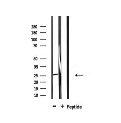
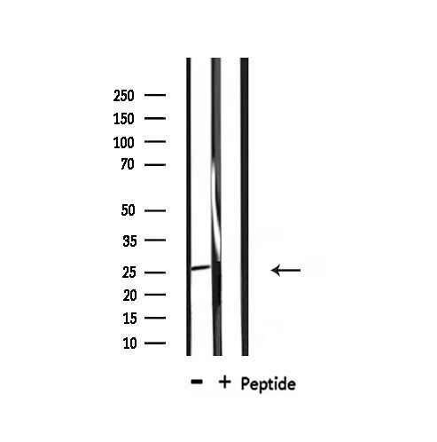
- Western blot analysis of CHOP in mouse brain. Samples were incubated with CHOP polyclonal antibody (Product # PA5-104528).
Supportive validation
- Submitted by
- Invitrogen Antibodies (provider)
- Main image
- Experimental details
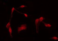
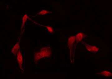
- Immunofluorescent analysis of CHOP in HeLa cells. Samples were fixed with paraformaldehyde, permeabilized with 0.1% Triton X-100, blocked with 10% serum (45 min at 25°C) incubated with CHOP polyclonal antibody (Product # PA5-104528) using a dilution of 1:200 (1 hr, 37°C), and followed by goat anti-rabbit IgG Alexa Fluor 594 at a dilution of 1:600.

- Submitted by
- Invitrogen Antibodies (provider)
- Main image

- Experimental details
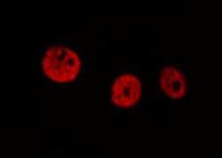
- Immunofluorescent analysis of CHOP in HeLa cells. Samples were fixed with paraformaldehyde, permeabilized with 0.1% Triton X-100, blocked with 10% serum (45 min at 25°C) incubated with CHOP polyclonal antibody (Product # PA5-104528) using a dilution of 1:200 (1 hr, 37°C), and followed by goat anti-rabbit IgG Alexa Fluor 594 at a dilution of 1:600.
- Submitted by
- Invitrogen Antibodies (provider)
- Main image
- Experimental details
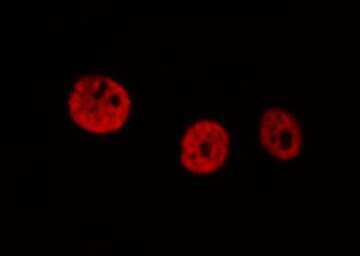
- Immunofluorescent analysis of CHOP in HeLa cells. Samples were fixed with paraformaldehyde, permeabilized with 0.1% Triton X-100, blocked with 10% serum (45 min at 25°C) incubated with CHOP polyclonal antibody (Product # PA5-104528) using a dilution of 1:200 (1 hr, 37°C), and followed by goat anti-rabbit IgG Alexa Fluor 594 at a dilution of 1:600.
Supportive validation
- Submitted by
- Invitrogen Antibodies (provider)
- Main image
- Experimental details
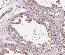
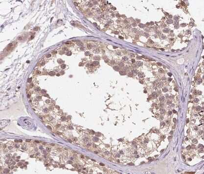
- Immunohistochemistry analysis of paraffin-embedded CHOP in human testis tissue sections. Antigen retrieval was performed using citrate buffer. Samples were blocked with blocking buffer (1.5 hr, 22°C), incubated with CHOP polyclonal antibody (Product # PA5-104528) using a dilution of 1:100 (1.5 hr, 22°C), followed by HRP conjugated goat anti-rabbit.
- Submitted by
- Invitrogen Antibodies (provider)
- Main image
- Experimental details
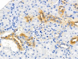
- Immunohistochemistry analysis of CHOP in rat kidney tissue. The sample was formaldehyde fixed and a heat mediated antigen retrieval step in citrate buffer was performed. Samples were incubated with CHOP polyclonal antibody (Product # PA5-104528) using a dilution of 1:100 (4°C overnight) followed by HRP conjugated anti-Rabbit secondary antibody.

- Submitted by
- Invitrogen Antibodies (provider)
- Main image

- Experimental details
- Immunohistochemistry analysis of paraffin-embedded CHOP in human testis tissue sections. Antigen retrieval was performed using citrate buffer. Samples were blocked with blocking buffer (1.5 hr, 22°C), incubated with CHOP polyclonal antibody (Product # PA5-104528) using a dilution of 1:100 (1.5 hr, 22°C), followed by HRP conjugated goat anti-rabbit.

Supportive validation
- Submitted by
- Invitrogen Antibodies (provider)
- Main image
- Experimental details
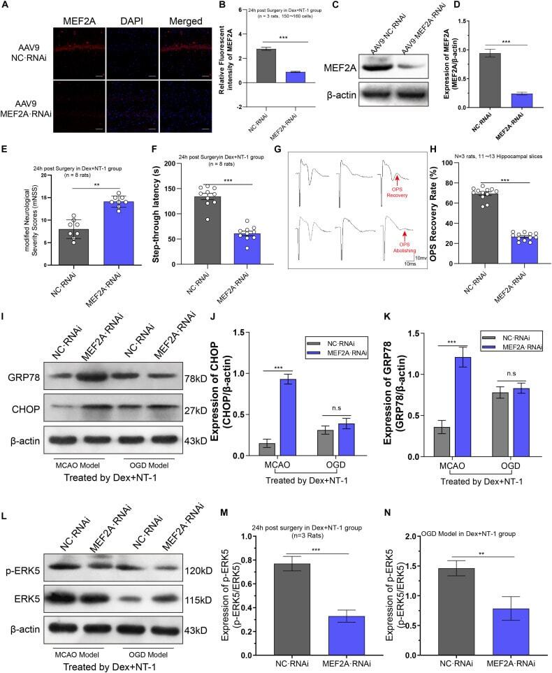
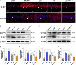
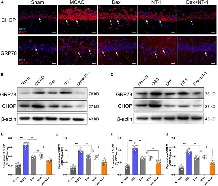
- FIGURE 5 Effects of dexmedetomidine and Netrin-1 on ERS protein (CHOP and GRP78) expression after MCAO and OGD injury. (A) CHOP (first line) and GRP78 (second line) expressions in hippocampus after MCAO injury. Immunofluorescence staining exhibited positive cells of CHOP and GRP78. Red indicated CHOP and GRP78 expression in plasm of pyramidal neurons, and blue indicated the nucleus. Scale bar = 50 mum. (B,C) Western blot analysis of ERS-related proteins CHOP and GRP78, beta-actin was used as an internal control. (D-G) Quantitative analysis of CHOP and GRP78 expression levels. * P < 0.05, ** P < 0.01 and *** P < 0.001 using one-way ANOVA followed by Tukey's post hoc tests.
- Submitted by
- Invitrogen Antibodies (provider)
- Main image
- Experimental details
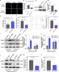
- FIGURE 7 Knockdown MEF2A weakened the therapeutic effect of dexmedetomidine combined with Netrin-1 after MCAO and OGD injuries. (A,B) Immunofluorescence staining exhibited downregulation in MEF2A expression in the hippocampus at 14 days after AAV9-MEF2A RNAi microinjection. (C,D) Western blot analysis shows downregulation in MEF2A expression in the hippocampus at 14 days after AAV9-MEF2A RNAi microinjection. (E) The mNSS result after MEF2A knockdown. (F) The step-through latency result after MEF2A knockdown. (G,H) The OPS recovery rate after MEF2A knockdown. (I-K) Western blot analysis of ERS-related proteins CHOP and GRP78 in the hippocampus at 14 days after AAV9-MEF2A RNAi microinjection, beta-actin was used as an internal control. (L-N) Western blot analysis of ERS-related proteins ERK5 and p-ERK5 in the hippocampus at 14 days after AAV9-MEF2A RNAi microinjection, beta-actin was used as an internal control. ** P < 0.01 and *** P < 0.001 using one-way ANOVA followed by Tukey's post hoc tests.