Explore
 Validate
 Learn
Learn Western blot
 ELISA
ELISAAntibody data
- Antibody Data
- Antigen structure
- References [3]
- Comments [0]
- Validations
- Western blot [1]
Submit
Validation data
Reference
Comment
Report error
- Product number
- A00311-2 - Provider product page

- Provider
- Boster Biological Technology
- Product name
- Anti-DDIT3 Antibody Picoband™
- Antibody type
- Polyclonal
- Description
- Rabbit IgG polyclonal antibody for DDIT3 detection. Tested with WB, IHC-P, FCM, Direct ELISA in Human.
- Reactivity
- Human
- Host
- Rabbit
- Vial size
- 100μg/vial
- Concentration
- Add 0.2ml of distilled water will yield a concentration of 500ug/ml.
- Storage
- At -20°C for one year. After reconstitution, at 4°C for one month. It can also be aliquoted and stored frozen at -20°C for a longer time. Avoid repeated freezing and thawing.
- Handling
- Add 0.2ml of distilled water will yield a concentration of 500ug/ml.
Submitted references Potential impacts of bisphenols on prostate cells: An overview of cytotoxicity, proliferation, oxidative stress, apoptosis, and ER-stress response activation.
Lycorine protects against septic myocardial injury by activating AMPK-related pathways.
Tension induces intervertebral disc degeneration via endoplasmic reticulum stress-mediated autophagy.
Caglayan M, Ozden S
Food and chemical toxicology : an international journal published for the British Industrial Biological Research Association 2024 Feb;184:114416
Food and chemical toxicology : an international journal published for the British Industrial Biological Research Association 2024 Feb;184:114416
Lycorine protects against septic myocardial injury by activating AMPK-related pathways.
Zhao H, Chen Y, Qian L, Du L, Wu X, Tian Y, Deng C, Liu S, Yang W, Lu C, Zhang Y, Ren J, Yang Y
Free radical biology & medicine 2023 Mar;197:1-14
Free radical biology & medicine 2023 Mar;197:1-14
Tension induces intervertebral disc degeneration via endoplasmic reticulum stress-mediated autophagy.
Chen J, Lin Z, Deng K, Shao B, Yang D
Bioscience reports 2019 Aug 30;39(8)
Bioscience reports 2019 Aug 30;39(8)
No comments: Submit comment
Supportive validation
- Submitted by
- Boster Biological Technology (provider)
- Main image

- Experimental details
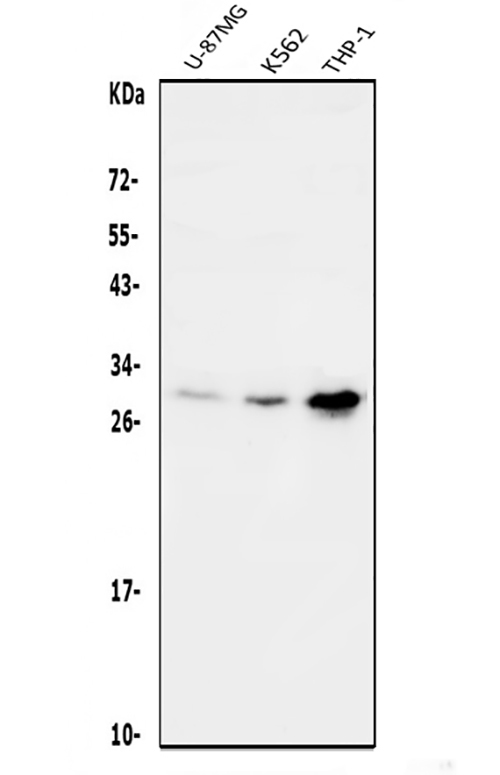
- Western blot analysis of DDIT3 using anti-DDIT3 antibody (A00311-2). Electrophoresis was performed on a 5-20% SDS-PAGE gel at 70V (Stacking gel) / 90V (Resolving gel) for 2-3 hours. The sample well of each lane was loaded with 50ug of sample under reducing conditions. Lane 1: human U-87MG whole cell lysates, Lane 2: human K562 whole cell lysates, Lane 3: human THP-1 whole cell lysates. After Electrophoresis, proteins were transferred to a Nitrocellulose membrane at 150mA for 50-90 minutes. Blocked the membrane with 5% Non-fat Milk/ TBS for 1.5 hour at RT. The membrane was incubated with rabbit anti-DDIT3 antigen affinity purified polyclonal antibody (Catalog # A00311-2) at 0.5 μg/mL overnight at 4°C, then washed with TBS-0.1%Tween 3 times with 5 minutes each and probed with a goat anti-rabbit IgG-HRP secondary antibody at a dilution of 1:5000 for 1.5 hour at RT. The signal is developed using an Enhanced Chemiluminescent detection (ECL) kit (Catalog # EK1002) with Tanon 5200 system. A specific band was detected for DDIT3 at approximately 29KD. The expected band size for DDIT3 is at 29KD.
- Additional image