 Explore
 Validate
 Learn
Learn Western blot
 Immunocytochemistry
Antibody data
- Antibody Data
- Antigen structure
- References [1]
- Comments [0]
- Validations
- Immunocytochemistry [2]
Submit
Validation data
Reference
Comment
Report error
- Product number
- PA5-28956 - Provider product page

- Provider
- Invitrogen Antibodies
- Product name
- CHOP Polyclonal Antibody
- Antibody type
- Polyclonal
- Antigen
- Recombinant full-length protein
- Description
- Recommended positive controls: HeLa, HeLa treated tunicaycin 8 hr. Predicted reactivity: Mouse (88%), Rat (90%), Pig (95%), Rhesus Monkey (98%), Bovine (92%). Store product as a concentrated solution. Centrifuge briefly prior to opening the vial.
- Reactivity
- Human
- Host
- Rabbit
- Isotype
- IgG
- Vial size
- 100 μL
- Concentration
- 1 mg/mL
- Storage
- Store at 4°C short term. For long term storage, store at -20°C, avoiding freeze/thaw cycles.
Submitted references Schizandrin A induces the apoptosis and suppresses the proliferation, invasion and migration of gastric cancer cells by activating endoplasmic reticulum stress.
Pu H, Qian Q, Wang F, Gong M, Ge X
Molecular medicine reports 2021 Nov;24(5)
Molecular medicine reports 2021 Nov;24(5)
No comments: Submit comment
Supportive validation
- Submitted by
- Invitrogen Antibodies (provider)
- Main image
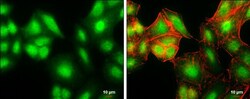
- Experimental details
- Immunocytochemistry-Immunofluorescence analysis of CHOP was performed in A549 cells fixed in 4% paraformaldehyde at RT for 15 min. Green: CHOP Polyclonal Antibody (Product # PA5-28956) diluted at 1:200. Red: phalloidin, a cytoskeleton marker. Scale bar = 10 µm.
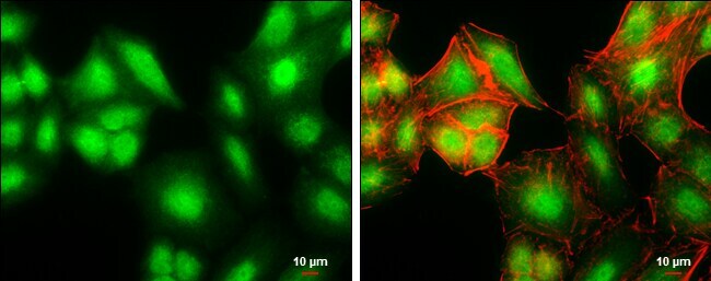
