Explore
 Validate
 Learn
Learn Western blot
 ELISA
ELISAAntibody data
- Antibody Data
- Antigen structure
- References [3]
- Comments [0]
- Validations
- Western blot [1]
- Flow cytometry [1]
Submit
Validation data
Reference
Comment
Report error
- Product number
- ABIN952424 - Provider product page

- Provider
- antibodies-online
- Product name
- anti-DNA-Damage-Inducible Transcript 3 (DDIT3) (C-Term), (AA 127-157) antibody
- Antibody type
- Polyclonal
- Antigen
- KLH conjugated synthetic peptide between 127-157 amino acids from the C-terminal region of Human DDIT3. Genename: DDIT3
- Description
- Peptide Affinity Chromatography on Protein A
- Reactivity
- Human, Mouse
- Host
- Rabbit
- Epitope
- C-Term,AA 127-157
- Vial size
- 0.4 mL
- Concentration
- 0.25 mg/mL
- Storage
- Store the antibody undiluted at 2-8°C for one month or (in aliquots) at -20°C for longer.
- Handling
- Avoid repeated freezing and thawing.
Submitted references Coxsackievirus B3 infection activates the unfolded protein response and induces apoptosis through downregulation of p58IPK and activation of CHOP and SREBP1.
Endoplasmic reticulum stress-induced transcription factor, CHOP, is crucial for dendritic cell IL-23 expression.
Repression of peroxisome proliferator-activated receptor gamma by mucosal ribotoxic insult-activated CCAAT/enhancer-binding protein homologous protein.
Zhang HM, Ye X, Su Y, Yuan J, Liu Z, Stein DA, Yang D
Journal of virology 2010 Sep;84(17):8446-59
Journal of virology 2010 Sep;84(17):8446-59
Endoplasmic reticulum stress-induced transcription factor, CHOP, is crucial for dendritic cell IL-23 expression.
Goodall JC, Wu C, Zhang Y, McNeill L, Ellis L, Saudek V, Gaston JS
Proceedings of the National Academy of Sciences of the United States of America 2010 Oct 12;107(41):17698-703
Proceedings of the National Academy of Sciences of the United States of America 2010 Oct 12;107(41):17698-703
Repression of peroxisome proliferator-activated receptor gamma by mucosal ribotoxic insult-activated CCAAT/enhancer-binding protein homologous protein.
Park SH, Choi HJ, Yang H, Do KH, Kim J, Moon Y
Journal of immunology (Baltimore, Md. : 1950) 2010 Nov 1;185(9):5522-30
Journal of immunology (Baltimore, Md. : 1950) 2010 Nov 1;185(9):5522-30
No comments: Submit comment
Supportive validation
- Submitted by
- antibodies-online (provider)
- Main image
- Experimental details
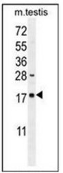
- Western blot analysis in Mouse testis tissue lysates (35ug/lane) using DDIT3 Antibody (C-term) Cat.-No AP51213PU-N. This demonstrates the DDIT3 antibody detected the DDIT3 protein (arrow).

Supportive validation
- Submitted by
- antibodies-online (provider)
- Main image

- Experimental details
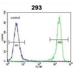
- Flow Cytometric analysis of 293 cells using DIT3 Antibody (C-term) Cat.-No AP51213PU-N (right histogram) compared to a Negative Control cell (left histogram). FITC-conjugated Goat-anti-Rabbit secondary antibodies were used for the analysis.