 Explore
 Validate
 Learn
Learn Western blot
Antibody data
- Antibody Data
- Antigen structure
- References [0]
- Comments [0]
- Validations
- Western blot [2]
- Immunocytochemistry [1]
- Immunohistochemistry [1]
- Flow cytometry [1]
Submit
Validation data
Reference
Comment
Report error
- Product number
- TA328618 - Provider product page

- Provider
- OriGene
- Product name
- Rabbit Polyclonal Anti-Human Protease-activated Receptor-1 (extracellular)
- Antibody type
- Polyclonal
- Description
- Rabbit Polyclonal Anti-Human Protease-activated Receptor-1 (extracellular)
- Host
- Rabbit
- Conjugate
- Unconjugated
- Epitope
- F2R
- Antibody clone number
- NULL
- Vial size
- 200 µl
- Concentration
- NULL
No comments: Submit comment
Supportive validation
- Submitted by
- OriGene (provider)
- Main image
- Experimental details
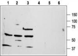
- Western blot analysis of human promyelocytic leukemia HL-60 (lanes 1 and 4), human T-cell leukemia Jurkat (lanes 2 and 5), and chronic myelogenous leukemia K562 (lanes 3 and 6) cell line lysates: 1-3. Anti-Human Protease-Activated Receptor-1 (extracellular) antibody, (1:200). 4-6. Anti-Human Protease-Activated Receptor-1 (extracellular) antibody, preincubated with the control peptide antigen.
- Validation comment
- WB

- Submitted by
- OriGene (provider)
- Main image

- Experimental details
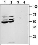
- Western blot analysis of human colon cancer HT-29 (lanes 1 and 3) and Colo-205 (lanes 2 and 4) cell line lysates: 1, 2. Anti-Human Protease-Activated Receptor-1 (extracellular) antibody, (1:200). 3, 4. Anti-Human Protease-Activated Receptor-1 (extracellular) antibody, preincubated with the control peptide antigen.
- Validation comment
- WB
Supportive validation
- Submitted by
- OriGene (provider)
- Main image
- Experimental details
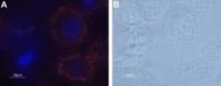
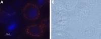
- Expression of PAR-1 in human prostate PC-3 cell line. Immunocytochemical staining of human prostate PC-3 cell line. A. Live intact PC-3 cells were stained with Anti-Human Protease-Activated Receptor-1 (extracellular) antibody, (1:50), followed by goat-anti-rabbit-AlexaFluor-555 secondary antibody (red staining). Nuclei were visualized using the cell-permeable dye Hoechst 33342 (blue). B. Live view of the same field as A.
- Validation comment
- IF
Supportive validation
- Submitted by
- OriGene (provider)
- Main image

- Experimental details
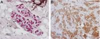
- IHC staining of paraffin-embedded human breast sections using Anti-Human Protease-Activated Receptor-1 (extracellular) antibody , (1:100). PAR-1 staining is highly specific for epithelium-derived cells. A. In the normal resting breast, epithelial cells of the mammary ducts are visible using Histofine (pink). B. The breast carcinoma contains epithelium-derived malignant cells stained with DAB (brown). Hematoxilin is used as the counterstain.
- Validation comment
- IHC
Supportive validation
- Submitted by
- OriGene (provider)
- Main image
- Experimental details
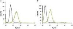
- Indirect Flow cytometry analysis of live intact HL-60 (human promyelocytic leukemia) (A) and Jurkat (human T cell leukemia) (B) cell lines: black line, Unstained Cells + FITC-conjugated goat anti-rabbit antibody. green line, Cells + Anti-Human Protease-Activated Receptor-1 (extracellular) antibody, (1:20) + FITC-conjugated goat anti-rabbit antibody.
- Validation comment
- FC
