Explore
 Validate
 Learn
Learn Western blot
Antibody data
- Antibody Data
- Antigen structure
- References [1]
- Comments [0]
- Validations
- Western blot [1]
- Immunohistochemistry [1]
- Other assay [2]
Submit
Validation data
Reference
Comment
Report error
- Product number
- PA5-94929 - Provider product page

- Provider
- Invitrogen Antibodies
- Product name
- PAR1 Polyclonal Antibody
- Antibody type
- Polyclonal
- Antigen
- Synthetic peptide
- Description
- Reconstitute with 0.2 mL of distilled water to yield a concentration of 500 µg/mL.
- Reactivity
- Human
- Host
- Rabbit
- Isotype
- IgG
- Vial size
- 100 µg
- Concentration
- 500 µg/mL
- Storage
- Store at 4°C short term. For long term storage, store at -20°C, avoiding freeze/thaw cycles.
Submitted references F2r negatively regulates osteoclastogenesis through inhibiting the Akt and NFκB signaling pathways.
Zhang Y, Wang H, Zhu G, Qian A, Chen W
International journal of biological sciences 2020;16(9):1629-1639
International journal of biological sciences 2020;16(9):1629-1639
No comments: Submit comment
Supportive validation
- Submitted by
- Invitrogen Antibodies (provider)
- Main image

- Experimental details
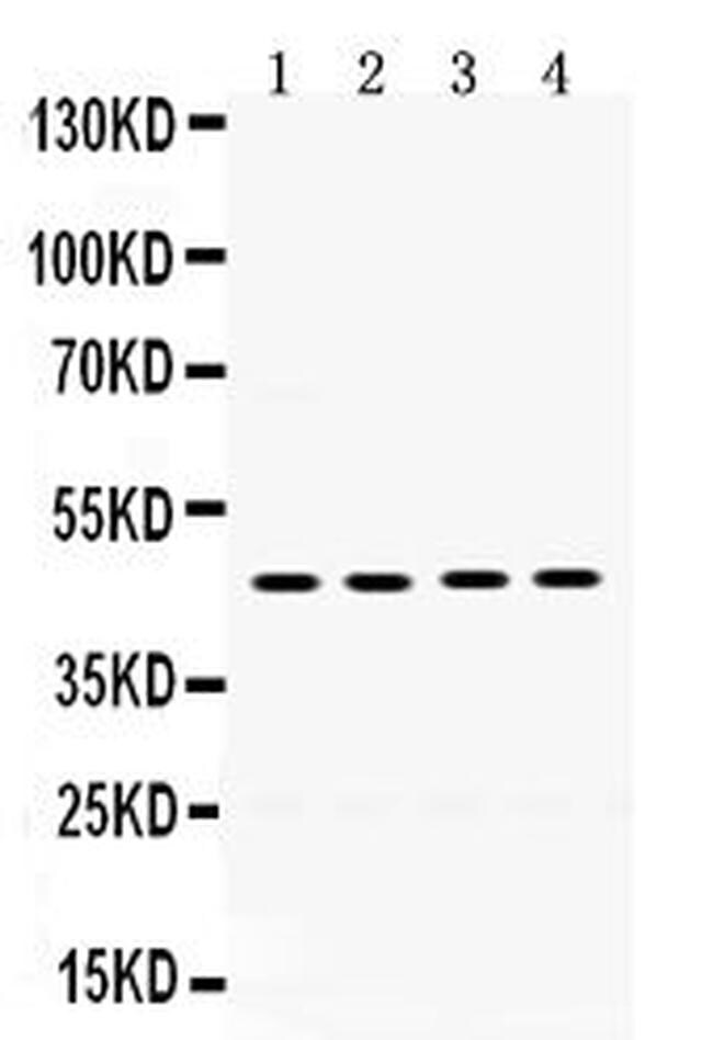
- Western blot analysis of PAR1 in Lane 1: MCF-7 whole cell lysate (40 µg), Lane 2: HeLa whole cell lysate (40 µg), Lane 3: 22RV1 whole cell lysate (40 µg), Lane 4: SW620 whole cell lysate (40 µg). Samples were incubated with PAR1 polyclonal antibody (Product # PA5-94929).
Supportive validation
- Submitted by
- Invitrogen Antibodies (provider)
- Main image

- Experimental details
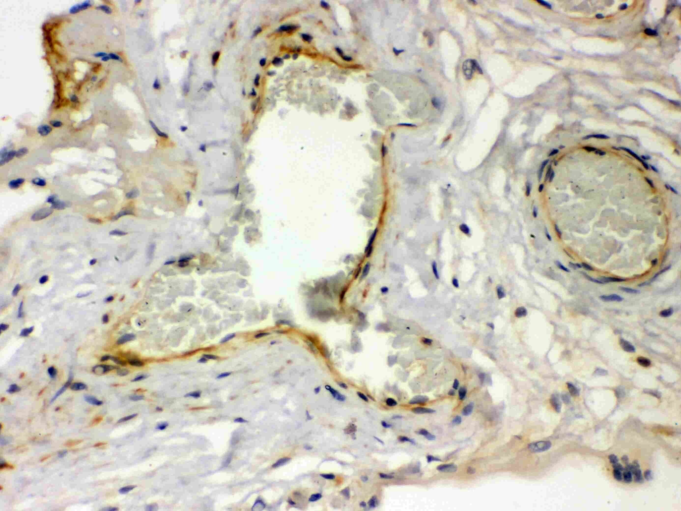
- Immunohistochemistry analysis of PAR1 in human placenta tissue. Samples were incubated with PAR1 polyclonal antibody (Product # PA5-94929).
Supportive validation
- Submitted by
- Invitrogen Antibodies (provider)
- Main image
- Experimental details
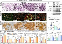
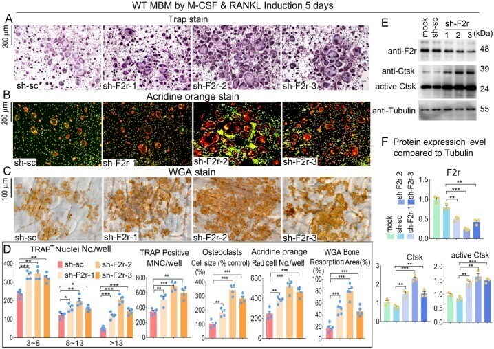
- Figure 2 F2r knockdown promotes osteoclastogenesis . ( A ) TRAP stain and ( B ) Acridine orange (AO) stain of sh-sc and sh-F2r infected MBMs that induced 5 days by M-CSF and RANKL in 12-well-plate. ( C ) Wheat germ agglutinin (WGA) stain of osteoclasts on bone slices to detect bone resorption area. ( D ) Quantification data of A-C . TRAP-positive MNCs (multinucleated cells, >=3 nuclei). ( E ) Western blot of F2r and Ctsk protein expression level in sh-sc and sh-F2r infected MBMs that induced 5 days by M-CSF and RANKL. ( F ) Quantification data of E by image J. Tubulin was used as a control. Protein expression level in the mock group was normalized as 1. Results are presented as mean +- SEM; one dot represents one sample. n>=3. ** p
- Submitted by
- Invitrogen Antibodies (provider)
- Main image
- Experimental details
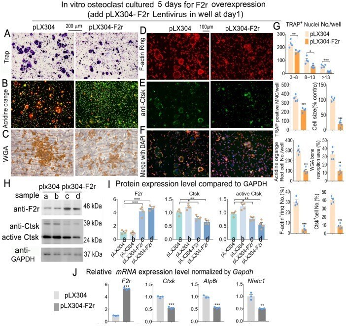
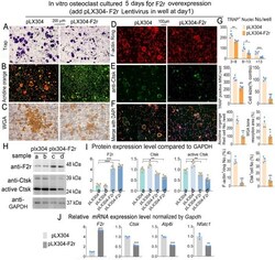
- Figure 3 F2r overexpression inhibited osteoclastogenesis. ( A ) TRAP stain and ( B ) Acridine orange stain of pLX304 and pLX304-F2r infected MBMs that induced 5 days by M-CSF and RANKL. ( C ) Wheat germ agglutinin (WGA) stain analysis of osteoclast on bone slices to detect bone resorption area of induced MBMs. ( D ) Fluorescence microscopy of F-actin ring stain and ( E ) anti-Ctsk immunofluorescence (IF) stain in mature osteoclasts. ( F ) Overlap of D, E was detected as a yellow-orange area in the merged image. ( G ) Quantification data of A-F. ( H ) Western blot of F2r and Ctsk protein expression level in pLX304 (a,b) and pLX304-F2r (c,d) infected osteoclasts. GAPDH was used as a control. Protein expression level in pLX304 group normalized as 1. ( I ) Quantification data of H. ( J ) qRT-PCR was carried out to measure F2r, Ctsk, Atp6i and Nfatc1 mRNA levels relative to Gapdh in osteoclasts. One dot represents one sample. Results are presented as mean +- SEM; n>=3. * p