Explore
 Validate
 Learn
Learn Western blot
 Immunocytochemistry
Antibody data
- Antibody Data
- Antigen structure
- References [2]
- Comments [0]
- Validations
- Immunocytochemistry [1]
- Flow cytometry [1]
Submit
Validation data
Reference
Comment
Report error
- Product number
- AF3855 - Provider product page

- Provider
- R&D Systems
- Product name
- Human PAR1 Antibody
- Antibody type
- Polyclonal
- Description
- Antigen Affinity-purified. Detects human PAR1 in direct ELISAs and Western blots.
- Reactivity
- Human
- Host
- Goat
- Conjugate
- Unconjugated
- Antigen sequence
P25116- Isotype
- IgG
- Vial size
- 100 ug
- Concentration
- LYOPH
- Storage
- Use a manual defrost freezer and avoid repeated freeze-thaw cycles. 12 months from date of receipt, -20 to -70 °C as supplied. 1 month, 2 to 8 °C under sterile conditions after reconstitution. 6 months, -20 to -70 °C under sterile conditions after reconstitution.
Submitted references An acidic microenvironment sets the humoral pattern recognition molecule PTX3 in a tissue repair mode.
Oncogenic epidermal growth factor receptor up-regulates multiple elements of the tissue factor signaling pathway in human glioma cells.
Doni A, Musso T, Morone D, Bastone A, Zambelli V, Sironi M, Castagnoli C, Cambieri I, Stravalaci M, Pasqualini F, Laface I, Valentino S, Tartari S, Ponzetta A, Maina V, Barbieri SS, Tremoli E, Catapano AL, Norata GD, Bottazzi B, Garlanda C, Mantovani A
The Journal of experimental medicine 2015 Jun 1;212(6):905-25
The Journal of experimental medicine 2015 Jun 1;212(6):905-25
Oncogenic epidermal growth factor receptor up-regulates multiple elements of the tissue factor signaling pathway in human glioma cells.
Magnus N, Garnier D, Rak J
Blood 2010 Aug 5;116(5):815-8
Blood 2010 Aug 5;116(5):815-8
No comments: Submit comment
Supportive validation
- Submitted by
- R&D Systems (provider)
- Main image
- Experimental details
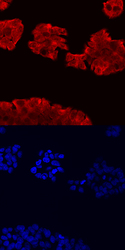
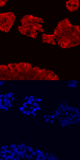
- PAR1 in HT-29 Human Cell Line. PAR1 was detected in immersion fixed HT-29 human colon adenocarcinoma cell line using Goat Anti-Human PAR1 Antigen Affinity-purified Polyclonal Antibody (Catalog # AF3855) at 10 µg/mL for 3 hours at room temperature. Cells were stained using the NorthernLights™ 557-conjugated Anti-Goat IgG Secondary Antibody (red, upper panel; Catalog # NL001) and counterstained with DAPI (blue, lower panel). Specific staining was localized to cytoplasm. View our protocol for Fluorescent ICC Staining of Cells on Coverslips.
Supportive validation
- Submitted by
- R&D Systems (provider)
- Main image
- Experimental details
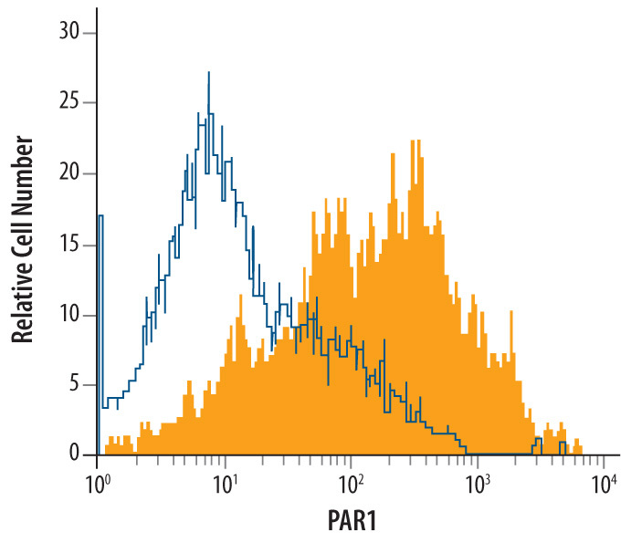
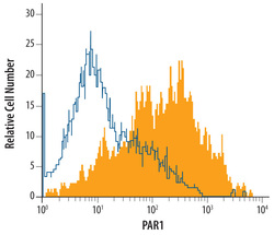
- Detection of PAR1 in HT-29 Human Cell Line by Flow Cytometry. HT-29 human colon adenocarcinoma cell line was stained with Human PAR1 Antigen Affinity-purified Polyclonal Antibody (Catalog # AF3855, filled histogram) or isotype control antibody (Catalog # AB-108-C, open histogram), followed by Phycoerythrin-conjugated Anti-Goat IgG Secondary Antibody (Catalog # F0108).