Explore
 Validate
 Learn
Learn Western blot
 Immunocytochemistry
Antibody data
- Antibody Data
- Antigen structure
- References [2]
- Comments [0]
- Validations
- Immunocytochemistry [2]
Submit
Validation data
Reference
Comment
Report error
- Product number
- PA1-753 - Provider product page

- Provider
- Invitrogen Antibodies
- Product name
- BACE2 Polyclonal Antibody
- Antibody type
- Polyclonal
- Antigen
- Synthetic peptide
- Description
- PA1-753 detects human beta-secretase 2 (BACE2) from transfected cells and rat and mouse samples. PA1-753 has been successfully used in Western blot and ICC/IF procedures. By Western blot this antibody detects an ~58 kDa protein representing BACE2 from SY-SY5Y cells overexpressing the human gene. The PA1-753 immunizing peptide corresponds to amino acid residues 441-457 from human BACE2. This sequence is completely conserved in mouse. The PA1-753 immunizing peptide (Cat. # PEP-166) is available for use in neutralization and control experiments.
- Reactivity
- Human, Mouse, Rat
- Host
- Rabbit
- Isotype
- IgG
- Vial size
- 100 μg
- Concentration
- 1 mg/mL
- Storage
- -20°C, Avoid Freeze/Thaw Cycles
Submitted references Interaction between amyloid precursor protein and Nogo receptors regulates amyloid deposition.
Identification of a novel aspartic protease (Asp 2) as beta-secretase.
Zhou X, Hu X, He W, Tang X, Shi Q, Zhang Z, Yan R
FASEB journal : official publication of the Federation of American Societies for Experimental Biology 2011 Sep;25(9):3146-56
FASEB journal : official publication of the Federation of American Societies for Experimental Biology 2011 Sep;25(9):3146-56
Identification of a novel aspartic protease (Asp 2) as beta-secretase.
Hussain I, Powell D, Howlett DR, Tew DG, Meek TD, Chapman C, Gloger IS, Murphy KE, Southan CD, Ryan DM, Smith TS, Simmons DL, Walsh FS, Dingwall C, Christie G
Molecular and cellular neurosciences 1999 Dec;14(6):419-27
Molecular and cellular neurosciences 1999 Dec;14(6):419-27
No comments: Submit comment
Supportive validation
- Submitted by
- Invitrogen Antibodies (provider)
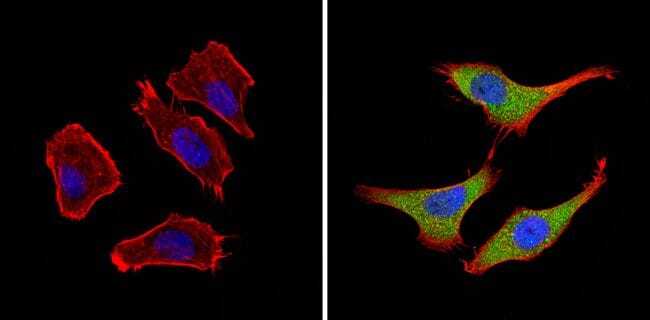
- Main image

- Experimental details
- Immunofluorescent analysis of BACE2 (green) showing staining in the nucleus and cytoplasm of A549 cells (right) compared to a negative control without primary antibody (left). Formalin-fixed cells were permeabilized with 0.1% Triton X-100 in TBS for 5-10 minutes and blocked with 3% BSA-PBS for 30 minutes at room temperature. Cells were probed with a BACE2 polyclonal antibody (Product # PA1-753) in 3% BSA-PBS at a dilution of 1:100 and incubated overnight at 4 ºC in a humidified chamber. Cells were washed with PBST and incubated with a DyLight-conjugated secondary antibody in PBS at room temperature in the dark. F-actin (red) was stained with a fluorescent red phalloidin and nuclei (blue) were stained with Hoechst or DAPI. Images were taken at a magnification of 60x.
- Submitted by
- Invitrogen Antibodies (provider)
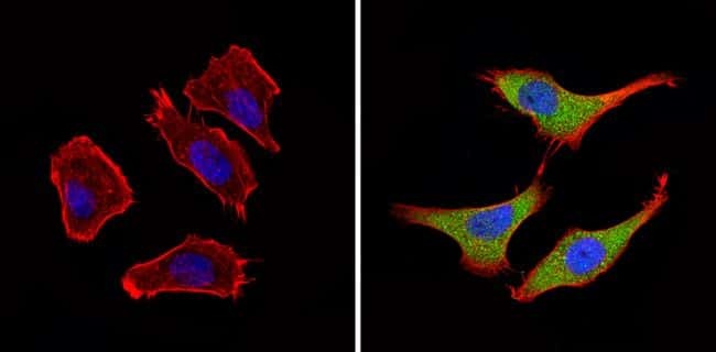
- Main image

- Experimental details
- Immunofluorescent analysis of BACE2 (green) showing staining in the nucleus and cytoplasm of A549 cells (right) compared to a negative control without primary antibody (left). Formalin-fixed cells were permeabilized with 0.1% Triton X-100 in TBS for 5-10 minutes and blocked with 3% BSA-PBS for 30 minutes at room temperature. Cells were probed with a BACE2 polyclonal antibody (Product # PA1-753) in 3% BSA-PBS at a dilution of 1:100 and incubated overnight at 4 ºC in a humidified chamber. Cells were washed with PBST and incubated with a DyLight-conjugated secondary antibody in PBS at room temperature in the dark. F-actin (red) was stained with a fluorescent red phalloidin and nuclei (blue) were stained with Hoechst or DAPI. Images were taken at a magnification of 60x.