 Explore
 Validate
 Learn
Learn Western blot
 Immunocytochemistry
Antibody data
- Antibody Data
- Antigen structure
- References [0]
- Comments [0]
- Validations
- Western blot [1]
- Immunocytochemistry [2]
- Immunohistochemistry [20]
Submit
Validation data
Reference
Comment
Report error
- Product number
- LS-C782188 - Provider product page

- Provider
- LSBio
- Product name
- CTTN / Cortactin Antibody LS-C782188
- Antibody type
- Polyclonal
- Description
- Antigen Affinity purification
- Reactivity
- Human, Mouse, Rat
- Host
- Rabbit
- Isotype
- IgG
- Storage
- After reconstitution, store at 4°C for up to 1 month. Long-term: aliquot and store at -20°C. Avoid freeze-thaws cycles.
No comments: Submit comment
Enhanced validation
- Submitted by
- LSBio (provider)
- Enhanced method
- Genetic validation
- Main image

- Experimental details
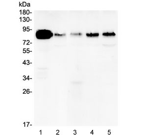
- Western blot testing of human 1) A431, 2) U-87 MG, 3) A549, 4) PC-3 and 5) HeLa lysate with Cortactin antibody at 0.5ug/ml. Predicted molecular weight ~61 kDa but routinely observed at ~80 kDa.
Supportive validation
- Submitted by
- LSBio (provider)
- Enhanced method
- Genetic validation
- Main image

- Experimental details
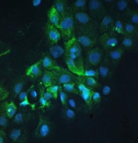
- IF/ICC staining of FFPE human A431 cells with Cortactin antibody at 1ug/ml. HIER: boil tissue sections in pH6, 10mM citrate buffer, for 10-20 min followed by cooling at RT for 20 min.
- Submitted by
- LSBio (provider)
- Main image

- Experimental details
- IF/ICC staining of FFPE human A431 cells with Cortactin antibody at 1ug/ml. HIER: boil tissue sections in pH6, 10mM citrate buffer, for 10-20 min followed by cooling at RT for 20 min.

Enhanced validation
- Submitted by
- LSBio (provider)
- Enhanced method
- Genetic validation
- Main image
- Experimental details
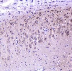
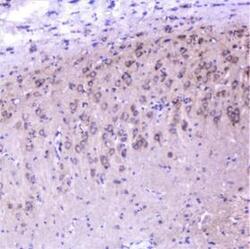
- IHC staining of FFPE rat brain with Cortactin antibody at 1ug/ml. HIER: boil tissue sections in pH6, 10mM citrate buffer, for 10-20 min followed by cooling at RT for 20 min.

- Submitted by
- LSBio (provider)
- Enhanced method
- Genetic validation
- Main image

- Experimental details
- IHC staining of FFPE rat brain with Cortactin antibody at 1ug/ml. HIER: boil tissue sections in pH6, 10mM citrate buffer, for 10-20 min followed by cooling at RT for 20 min.

- Submitted by
- LSBio (provider)
- Enhanced method
- Genetic validation
- Main image

- Experimental details
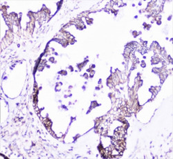
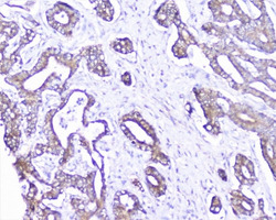
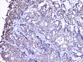
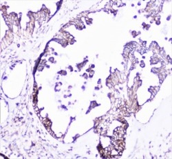
- IHC staining of FFPE human gastric carcinoma with Cortactin antibody at 1ug/ml. HIER: boil tissue sections in pH6, 10mM citrate buffer, for 10-20 min followed by cooling at RT for 20 min.

- Submitted by
- LSBio (provider)
- Enhanced method
- Genetic validation
- Main image
- Experimental details
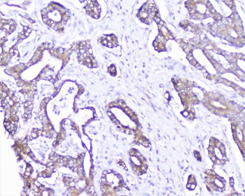
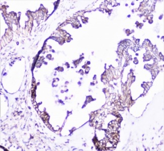
- IHC staining of FFPE human ovarian carcinoma with Cortactin antibody at 1ug/ml. HIER: boil tissue sections in pH6, 10mM citrate buffer, for 10-20 min followed by cooling at RT for 20 min.

- Submitted by
- LSBio (provider)
- Enhanced method
- Genetic validation
- Main image
- Experimental details
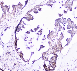
- IHC staining of FFPE human cholangiocarcinoma carcinoma with Cortactin antibody at 1ug/ml. HIER: boil tissue sections in pH6, 10mM citrate buffer, for 10-20 min followed by cooling at RT for 20 min.
- Submitted by
- LSBio (provider)
- Enhanced method
- Genetic validation
- Main image

- Experimental details
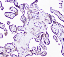
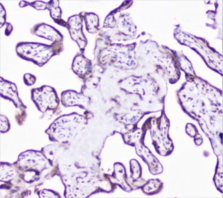
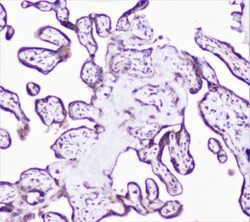
- IHC staining of FFPE human placenta with Cortactin antibody at 1ug/ml. HIER: boil tissue sections in pH6, 10mM citrate buffer, for 10-20 min followed by cooling at RT for 20 min.

- Submitted by
- LSBio (provider)
- Enhanced method
- Genetic validation
- Main image

- Experimental details
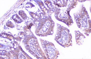
- IHC staining of FFPE mouse intestine with Cortactin antibody at 1ug/ml. HIER: boil tissue sections in pH6, 10mM citrate buffer, for 10-20 min followed by cooling at RT for 20 min.

- Submitted by
- LSBio (provider)
- Enhanced method
- Genetic validation
- Main image

- Experimental details
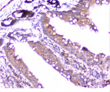
- IHC staining of FFPE rat intestine with Cortactin antibody at 1ug/ml. HIER: boil tissue sections in pH6, 10mM citrate buffer, for 10-20 min followed by cooling at RT for 20 min.

- Submitted by
- LSBio (provider)
- Enhanced method
- Genetic validation
- Main image

- Experimental details
- IHC staining of FFPE human gastric carcinoma with Cortactin antibody at 1ug/ml. HIER: boil tissue sections in pH6, 10mM citrate buffer, for 10-20 min followed by cooling at RT for 20 min.

- Submitted by
- LSBio (provider)
- Enhanced method
- Genetic validation
- Main image

- Experimental details
- IHC staining of FFPE human ovarian carcinoma with Cortactin antibody at 1ug/ml. HIER: boil tissue sections in pH6, 10mM citrate buffer, for 10-20 min followed by cooling at RT for 20 min.
- Submitted by
- LSBio (provider)
- Enhanced method
- Genetic validation
- Main image
- Experimental details
- IHC staining of FFPE human placenta with Cortactin antibody at 1ug/ml. HIER: boil tissue sections in pH6, 10mM citrate buffer, for 10-20 min followed by cooling at RT for 20 min.
- Submitted by
- LSBio (provider)
- Enhanced method
- Genetic validation
- Main image

- Experimental details
- IHC staining of FFPE mouse intestine with Cortactin antibody at 1ug/ml. HIER: boil tissue sections in pH6, 10mM citrate buffer, for 10-20 min followed by cooling at RT for 20 min.
- Submitted by
- LSBio (provider)
- Enhanced method
- Genetic validation
- Main image

- Experimental details
- IHC staining of FFPE rat intestine with Cortactin antibody at 1ug/ml. HIER: boil tissue sections in pH6, 10mM citrate buffer, for 10-20 min followed by cooling at RT for 20 min.
- Submitted by
- LSBio (provider)
- Main image

- Experimental details
- IHC staining of FFPE human gastric carcinoma with Cortactin antibody at 1ug/ml. HIER: boil tissue sections in pH6, 10mM citrate buffer, for 10-20 min followed by cooling at RT for 20 min.
- Submitted by
- LSBio (provider)
- Main image
- Experimental details
- IHC staining of FFPE human ovarian carcinoma with Cortactin antibody at 1ug/ml. HIER: boil tissue sections in pH6, 10mM citrate buffer, for 10-20 min followed by cooling at RT for 20 min.
- Submitted by
- LSBio (provider)
- Main image

- Experimental details
- IHC staining of FFPE human cholangiocarcinoma carcinoma with Cortactin antibody at 1ug/ml. HIER: boil tissue sections in pH6, 10mM citrate buffer, for 10-20 min followed by cooling at RT for 20 min.

- Submitted by
- LSBio (provider)
- Main image
- Experimental details
- IHC staining of FFPE human placenta with Cortactin antibody at 1ug/ml. HIER: boil tissue sections in pH6, 10mM citrate buffer, for 10-20 min followed by cooling at RT for 20 min.

- Submitted by
- LSBio (provider)
- Main image

- Experimental details
- IHC staining of FFPE mouse intestine with Cortactin antibody at 1ug/ml. HIER: boil tissue sections in pH6, 10mM citrate buffer, for 10-20 min followed by cooling at RT for 20 min.

- Submitted by
- LSBio (provider)
- Main image

- Experimental details
- IHC staining of FFPE rat intestine with Cortactin antibody at 1ug/ml. HIER: boil tissue sections in pH6, 10mM citrate buffer, for 10-20 min followed by cooling at RT for 20 min.

- Submitted by
- LSBio (provider)
- Main image
- Experimental details
- IHC staining of FFPE rat brain with Cortactin antibody at 1ug/ml. HIER: boil tissue sections in pH6, 10mM citrate buffer, for 10-20 min followed by cooling at RT for 20 min.
