 Explore
 Validate
 Learn
Learn Western blot
 Immunocytochemistry
Antibody data
- Antibody Data
- Antigen structure
- References [1]
- Comments [0]
- Validations
- Immunocytochemistry [2]
- Immunohistochemistry [2]
- Other assay [2]
Submit
Validation data
Reference
Comment
Report error
- Product number
- PA5-27134 - Provider product page

- Provider
- Invitrogen Antibodies
- Product name
- Cortactin Polyclonal Antibody
- Antibody type
- Polyclonal
- Antigen
- Synthetic peptide
- Description
- Recommended positive controls: HeLa, HepG2, NIH-3T3, JC, PC-12, mouse brain, rat brain. Predicted reactivity: Rhesus Monkey (100%). Store product as a concentrated solution. Centrifuge briefly prior to opening the vial.
- Reactivity
- Human, Mouse, Rat
- Host
- Rabbit
- Isotype
- IgG
- Vial size
- 100 μL
- Concentration
- 1 mg/mL
- Storage
- Store at 4°C short term. For long term storage, store at -20°C, avoiding freeze/thaw cycles.
Submitted references Mitochondrial mass governs the extent of human T cell senescence.
Callender LA, Carroll EC, Bober EA, Akbar AN, Solito E, Henson SM
Aging cell 2020 Feb;19(2):e13067
Aging cell 2020 Feb;19(2):e13067
No comments: Submit comment
Supportive validation
- Submitted by
- Invitrogen Antibodies (provider)
- Main image

- Experimental details
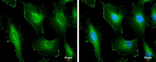
- Immunocytochemistry-Immunofluorescence analysis of Cortactin was performed in HeLa cells fixed in 4% paraformaldehyde at RT for 15 min. Green: Cortactin Polyclonal Antibody (Product # PA5-27134) diluted at 1:500. Blue: Hoechst 33342 staining. Scale bar = 10 µm.
- Submitted by
- Invitrogen Antibodies (provider)
- Main image
- Experimental details
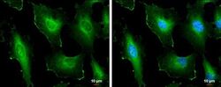
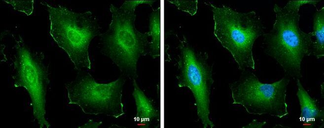
- Immunocytochemistry-Immunofluorescence analysis of Cortactin was performed in HeLa cells fixed in 4% paraformaldehyde at RT for 15 min. Green: Cortactin Polyclonal Antibody (Product # PA5-27134) diluted at 1:500. Blue: Hoechst 33342 staining. Scale bar = 10 µm.
Supportive validation
- Submitted by
- Invitrogen Antibodies (provider)
- Main image
- Experimental details
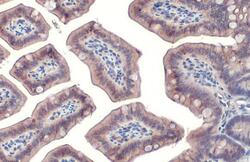
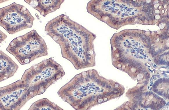
- Cortactin Polyclonal Antibody detects Cortactin protein at cell membrane and cytoplasm by immunohistochemical analysis. Sample: Paraffin-embedded mouse intestine. Cortactin stained by Cortactin Polyclonal Antibody (Product # PA5-27134) diluted at 1:500. Antigen Retrieval: Citrate buffer, pH 6.0, 15 min.
- Submitted by
- Invitrogen Antibodies (provider)
- Main image
- Experimental details
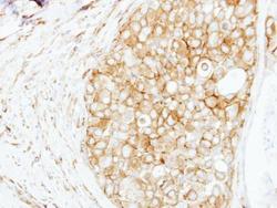
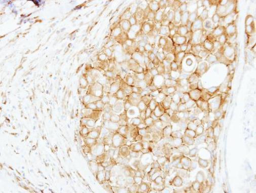
- Immunohistochemical analysis of paraffin-embedded human breast cancer, using Cortactin (Product # PA5-27134) antibody at 1:250 dilution. Antigen Retrieval: EDTA based buffer, pH 8.0, 15 min.
Supportive validation
- Submitted by
- Invitrogen Antibodies (provider)
- Main image
- Experimental details
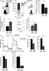
- Figure 2 Mitochondrial dysfunction is observed in CD8 + but not CD4 + EMRA T cell subsets. (a) Representative flow cytometry plots and cumulative graphs of TMRE staining from middle-aged donors showing membrane potential in CD45RA/CD27 T cell subsets directly ex vivo defined showing the percentage of cortactin-positive (a) CD4 + and (b) CD8 + T cells analysed directly ex vivo. Data expressed as mean +- SEM of six donors. (b) Mitochondrial ROS measured using MitoSOX by flow cytometry in CD4 + and CD8 + EMRA T cells from middle-aged donors. Data expressed as mean +- SEM of six donors. (c) Mitochondrial ROS production expressed as a ratio of mitochondrial mass. Calculated from data shown in Figures 1 b and 2 . (d) gammaH2AX expression as determined by flow cytometry in CD45RA/CD27-defined T cell subsets directly ex vivo from middle-aged donors; the graph shows the mean +- SEM for five donors. (e) Oxygen consumption rates (OCR) of the EMRA CD4 + and CD8 + T cell subsets from middle-aged donors were measured following a 15-min stimulation with 0.5 ug/ml anti-CD3 and 5 ng/ml IL-2; the cells were then subjected to a metabolic stress test using the indicated mitochondrial inhibitors. Data are representative of four independent experiments. (f) The basal OCR, extracellular acidification rate (ECAR) and spare respiratory capacity were measured following a 15-min stimulation with 0.5 ug/ml anti-CD3 and 5 ng/ml IL-2. Graphs show the mean +- SEM for four donors. (g) ATP concentration in E

- Submitted by
- Invitrogen Antibodies (provider)
- Main image
- Experimental details
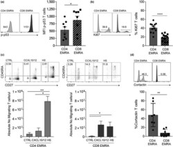
- Figure 4 Impaired function observed in CD8 + EMRA T cells. (a) Example and graph showing the expression of p-p53 in CD4 + and CD8 + CD45RA/CD27-defined EMRA T cells directly ex vivo from middle-aged donors. Graphs show the mean +- SEM for four donors. (b) Proliferation was defined in senescent T cell subsets using Ki67 directly ex vivo from middle-aged donors . Data show the mean +- SEM for 12 donors. (c) The migration of CD4 + and CD8 + EMRA T cells from middle-aged donors through HUVECs and their supporting transwell filters. HUVECs were stimulated with 20% decomplemented (heated at 56degC for 20 min) autologous donor sera for 24 hr. PBMCs were allowed to adhere and migrate for 4 hr towards either media, CXCL10/12 or autologous serum. The number of T cells was counted and expressed as a percentage of the total migrated CD4 + or CD8 + T cells. Data are expressed as the mean +- SEM of six donors. (d) Representative flow cytometry plots and cumulative graphs showing the percentage of cortactin-positive senescent T cell subsets analysed directly ex vivo from middle-aged donors. Data expressed as mean +- SEM of seven donors. p -values were calculated using a t test. * p < .05, ** p < .01, *** p < .005, and **** p < .001
