Explore
 Validate
 Learn
Learn Immunocytochemistry
 Flow cytometry
 Functional assay
Antibody data
- Antibody Data
- Antigen structure
- References [1]
- Comments [0]
- Validations
- Immunocytochemistry [4]
Submit
Validation data
Reference
Comment
Report error
- Product number
- ALX-804-232-C100 - Provider product page

- Provider
- Enzo Life Sciences
- Proper citation
- Enzo Life Sciences Cat#ALX-804-232-C100, RRID:AB_2051391
- Product name
- FasL (human) monoclonal antibody (2C101)
- Antibody type
- Monoclonal
- Antigen
- Recombinant protein fragment
- Description
- Affinity purified.
- Reactivity
- Human
- Host
- Mouse
- Isotype
- IgG
- Antibody clone number
- 2C101
- Vial size
- 100 μg
- Storage
- +4°C
- Handling
- Keep sterile. Do not freeze.
Submitted references IL-10 induces apoptosis in human monocytes involving the CD95 receptor/ligand pathway.
Schmidt M, Lügering N, Pauels HG, Schulze-Osthoff K, Domschke W, Kucharzik T
European journal of immunology 2000 Jun;30(6):1769-77
European journal of immunology 2000 Jun;30(6):1769-77
No comments: Submit comment
Supportive validation
- Submitted by
- Enzo Life Sciences (provider)
- Main image
- Experimental details
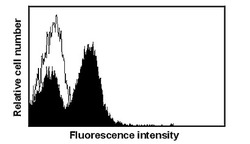
- Flow cytometric detection of FasL expression in stimulated H9 T-cells. Method: H9 T-cells (1x10E6) were either left untreated (open histogram) or stimulated with 10ng/ml PMA and 1µg/ml ionomycin (filled histogram). After 12 hours cells were harvested and stained with anti-FasL 2C101 (1µg/ml), followed by biotinylated goat anti-mouse IgG and streptavidin-PE.

- Submitted by
- Enzo Life Sciences (provider)
- Main image

- Experimental details
- Inhibition of apoptosis in anti-CD3 stimulated Jurkat T-cells by anti-Fas L. Method: Jurkat JR cells (2x10E6) were stimulated with plate-bound anti-CD3 antibodies (10µg/ml) in the presence or absence of the indicated concentrations of anti-FasL 2C101 or a control IgG1 mouse monoclonal antibody. After 48 hours apoptosis was evaluated by flow cytometric analysis of PI-stained nuclei.

- Submitted by
- Enzo Life Sciences (provider)
- Main image

- Experimental details
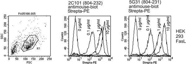
- Figures: Anti-FasL MAb ALX-804-232 on HEK 293 wt. and HEK 293-FasL clone cells expressing FasL fusion protein. HEK 293 cells transfected with membrane bound FasL (right figure) were compared with HEK 293 untransfected control cells (left figure).

- Submitted by
- Enzo Life Sciences (provider)
- Main image

- Experimental details
- Comparison of anti-FasL MAbs 804-231 and 804-232 on 293-FasL clone cells expressing FasL fusion protein. Comparable results were obtained for all other MAbs to hFasL recommended for flow cytometry.