 Explore
 Validate
 Learn
LearnPA3-305
antibody from Invitrogen Antibodies
Targeting: CXCR4
CD184, D2S201E, fusin, HM89, HSY3RR, LESTR, NPY3R, NPYR, NPYY3R
 Western blot
 Immunocytochemistry
Antibody data
- Antibody Data
- Antigen structure
- References [13]
- Comments [0]
- Validations
- Immunocytochemistry [4]
- Immunohistochemistry [1]
- Flow cytometry [2]
- Other assay [8]
Submit
Validation data
Reference
Comment
Report error
- Product number
- PA3-305 - Provider product page

- Provider
- Invitrogen Antibodies
- Product name
- CXCR4 Polyclonal Antibody
- Antibody type
- Polyclonal
- Antigen
- Synthetic peptide
- Description
- Several different molecular weight isoforms of CXCR4 have been described in the literature, which vary depending on cell type tested and extraction buffer used. Using Ramos, Raji, and Jurkat membrane fractions, two different MW forms of CXCR4 were detected with PA3-305 by Western blot, between 55-72kD. It is recommended to use a membrane isolation buffer/kit for extracting CXCR4 for Western blot analysis. Cellular membrane fractions were isolated for Western blot using Mem-PER Plus (Product # 89842).
- Reactivity
- Human, Mouse
- Host
- Rabbit
- Isotype
- IgG
- Vial size
- 100 μL
- Concentration
- Conc. Not Determined
- Storage
- -20°C, Avoid Freeze/Thaw Cycles
Submitted references Lidocaine inhibited migration of NSCLCA549 cells via the CXCR4 regulation.
Enhanced anti-inflammatory effects of mesenchymal stromal cells mediated by the transient ectopic expression of CXCR4 and IL10.
CXCL12 enhances pregnancy outcome via improvement of endometrial receptivity in mice.
CXCL12-CXCR4 Interplay Facilitates Palatal Osteogenesis in Mice.
An injectable hydrogel-formulated inhibitor of prolyl-4-hydroxylase promotes T regulatory cell recruitment and enhances alveolar bone regeneration during resolution of experimental periodontitis.
Oral lichen planus interactome reveals CXCR4 and CXCL12 as candidate therapeutic targets.
CXCR4 or CXCR7 antagonists treat endometriosis by reducing bone marrow cell trafficking.
Prognostic value of immunoexpression of CCR4, CCR5, CCR7 and CXCR4 in squamous cell carcinoma of tongue and floor of the mouth.
CXCR4 receptor blockage reduces the contribution of tumor and stromal cells to the metastatic growth in the liver.
Locoregional Confinement and Major Clinical Benefit of (188)Re-Loaded CXCR4-Targeted Nanocarriers in an Orthotopic Human to Mouse Model of Glioblastoma.
CXCR4 regulates interneuron migration in the developing neocortex.
CXCR4 regulates interneuron migration in the developing neocortex.
A dual role for the SDF-1/CXCR4 chemokine receptor system in adult brain: isoform-selective regulation of SDF-1 expression modulates CXCR4-dependent neuronal plasticity and cerebral leukocyte recruitment after focal ischemia.
Xing B, Yang L, Cui Y
Cancer biomarkers : section A of Disease markers 2022;33(3):317-330
Cancer biomarkers : section A of Disease markers 2022;33(3):317-330
Enhanced anti-inflammatory effects of mesenchymal stromal cells mediated by the transient ectopic expression of CXCR4 and IL10.
Hervás-Salcedo R, Fernández-García M, Hernando-Rodríguez M, Quintana-Bustamante O, Segovia JC, Alvarez-Silva M, García-Arranz M, Minguez P, Del Pozo V, de Alba MR, García-Olmo D, Ayuso C, Lamana ML, Bueren JA, Yañez RM
Stem cell research & therapy 2021 Feb 12;12(1):124
Stem cell research & therapy 2021 Feb 12;12(1):124
CXCL12 enhances pregnancy outcome via improvement of endometrial receptivity in mice.
Koo HS, Yoon MJ, Hong SH, Ahn J, Cha H, Lee D, Ko JE, Kwon H, Choi DH, Lee KA, Ko JJ, Kang YJ
Scientific reports 2021 Apr 1;11(1):7397
Scientific reports 2021 Apr 1;11(1):7397
CXCL12-CXCR4 Interplay Facilitates Palatal Osteogenesis in Mice.
Verheijen N, Suttorp CM, van Rheden REM, Regan RF, Helmich MPAC, Kuijpers-Jagtman AM, Wagener FADTG
Frontiers in cell and developmental biology 2020;8:771
Frontiers in cell and developmental biology 2020;8:771
An injectable hydrogel-formulated inhibitor of prolyl-4-hydroxylase promotes T regulatory cell recruitment and enhances alveolar bone regeneration during resolution of experimental periodontitis.
Nagai K, Ideguchi H, Kajikawa T, Li X, Chavakis T, Cheng J, Messersmith PB, Heber-Katz E, Hajishengallis G
FASEB journal : official publication of the Federation of American Societies for Experimental Biology 2020 Oct;34(10):13726-13740
FASEB journal : official publication of the Federation of American Societies for Experimental Biology 2020 Oct;34(10):13726-13740
Oral lichen planus interactome reveals CXCR4 and CXCL12 as candidate therapeutic targets.
Rivera C, Crisóstomo MF, Peña C, González-Díaz P, González-Arriagada WA
Scientific reports 2020 Mar 25;10(1):5454
Scientific reports 2020 Mar 25;10(1):5454
CXCR4 or CXCR7 antagonists treat endometriosis by reducing bone marrow cell trafficking.
Pluchino N, Mamillapalli R, Shaikh S, Habata S, Tal A, Gaye M, Taylor HS
Journal of cellular and molecular medicine 2020 Feb;24(4):2464-2474
Journal of cellular and molecular medicine 2020 Feb;24(4):2464-2474
Prognostic value of immunoexpression of CCR4, CCR5, CCR7 and CXCR4 in squamous cell carcinoma of tongue and floor of the mouth.
Domingueti CB, Janini JB, Paranaíba LM, Lozano-Burgos C, Olivero P, González-Arriagada WA
Medicina oral, patologia oral y cirugia bucal 2019 May 1;24(3):e354-e363
Medicina oral, patologia oral y cirugia bucal 2019 May 1;24(3):e354-e363
CXCR4 receptor blockage reduces the contribution of tumor and stromal cells to the metastatic growth in the liver.
Benedicto A, Romayor I, Arteta B
Oncology reports 2018 Apr;39(4):2022-2030
Oncology reports 2018 Apr;39(4):2022-2030
Locoregional Confinement and Major Clinical Benefit of (188)Re-Loaded CXCR4-Targeted Nanocarriers in an Orthotopic Human to Mouse Model of Glioblastoma.
Séhédic D, Chourpa I, Tétaud C, Griveau A, Loussouarn C, Avril S, Legendre C, Lepareur N, Wion D, Hindré F, Davodeau F, Garcion E
Theranostics 2017;7(18):4517-4536
Theranostics 2017;7(18):4517-4536
CXCR4 regulates interneuron migration in the developing neocortex.
Stumm RK, Zhou C, Ara T, Lazarini F, Dubois-Dalcq M, Nagasawa T, Höllt V, Schulz S
The Journal of neuroscience : the official journal of the Society for Neuroscience 2003 Jun 15;23(12):5123-30
The Journal of neuroscience : the official journal of the Society for Neuroscience 2003 Jun 15;23(12):5123-30
A dual role for the SDF-1/CXCR4 chemokine receptor system in adult brain: isoform-selective regulation of SDF-1 expression modulates CXCR4-dependent neuronal plasticity and cerebral leukocyte recruitment after focal ischemia.
Stumm RK, Rummel J, Junker V, Culmsee C, Pfeiffer M, Krieglstein J, Höllt V, Schulz S
The Journal of neuroscience : the official journal of the Society for Neuroscience 2002 Jul 15;22(14):5865-78
The Journal of neuroscience : the official journal of the Society for Neuroscience 2002 Jul 15;22(14):5865-78
No comments: Submit comment
Supportive validation
- Submitted by
- Invitrogen Antibodies (provider)
- Main image
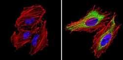
- Experimental details
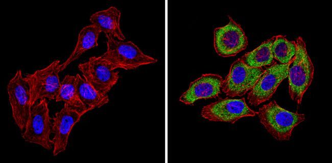
- Immunofluorescent analysis of CXCR4 (green) showing staining in the cytoplasm of Hela cells (right) compared to a negative control without primary antibody (left). Formalin-fixed cells were permeabilized with 0.1% Triton X-100 in TBS for 5-10 minutes and blocked with 3% BSA-PBS for 30 minutes at room temperature. Cells were probed with a CXCR4 polyclonal antibody (Product # PA3-305) in 3% BSA-PBS at a dilution of 1:100 and incubated overnight at 4ºC in a humidified chamber. Cells were washed with PBST and incubated with a DyLight-conjugated secondary antibody in PBS at room temperature in the dark. Actin was stained using Alexa Fluor 554 (red) and nuclei were stained with Hoechst or DAPI (blue). Images were taken at a magnification of 60x.

- Submitted by
- Invitrogen Antibodies (provider)
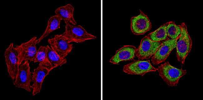
- Main image

- Experimental details
- Immunofluorescent analysis of CXCR4 (green) showing staining in the cytoplasm of U251 cells (right) compared to a negative control without primary antibody (left). Formalin-fixed cells were permeabilized with 0.1% Triton X-100 in TBS for 5-10 minutes and blocked with 3% BSA-PBS for 30 minutes at room temperature. Cells were probed with a CXCR4 polyclonal antibody (Product # PA3-305) in 3% BSA-PBS at a dilution of 1:100 and incubated overnight at 4ºC in a humidified chamber. Cells were washed with PBST and incubated with a DyLight-conjugated secondary antibody in PBS at room temperature in the dark. Actin was stained using Alexa Fluor 554 (red) and nuclei were stained with Hoechst or DAPI (blue). Images were taken at a magnification of 60x.
- Submitted by
- Invitrogen Antibodies (provider)
- Main image
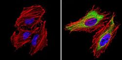
- Experimental details
- Immunofluorescent analysis of CXCR4 (green) showing staining in the cytoplasm of Hela cells (right) compared to a negative control without primary antibody (left). Formalin-fixed cells were permeabilized with 0.1% Triton X-100 in TBS for 5-10 minutes and blocked with 3% BSA-PBS for 30 minutes at room temperature. Cells were probed with a CXCR4 polyclonal antibody (Product # PA3-305) in 3% BSA-PBS at a dilution of 1:100 and incubated overnight at 4ºC in a humidified chamber. Cells were washed with PBST and incubated with a DyLight-conjugated secondary antibody in PBS at room temperature in the dark. Actin was stained using Alexa Fluor 554 (red) and nuclei were stained with Hoechst or DAPI (blue). Images were taken at a magnification of 60x.

- Submitted by
- Invitrogen Antibodies (provider)
- Main image
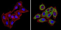
- Experimental details
- Immunofluorescent analysis of CXCR4 (green) showing staining in the cytoplasm of U251 cells (right) compared to a negative control without primary antibody (left). Formalin-fixed cells were permeabilized with 0.1% Triton X-100 in TBS for 5-10 minutes and blocked with 3% BSA-PBS for 30 minutes at room temperature. Cells were probed with a CXCR4 polyclonal antibody (Product # PA3-305) in 3% BSA-PBS at a dilution of 1:100 and incubated overnight at 4ºC in a humidified chamber. Cells were washed with PBST and incubated with a DyLight-conjugated secondary antibody in PBS at room temperature in the dark. Actin was stained using Alexa Fluor 554 (red) and nuclei were stained with Hoechst or DAPI (blue). Images were taken at a magnification of 60x.
Supportive validation
- Submitted by
- Invitrogen Antibodies (provider)
- Main image
- Experimental details
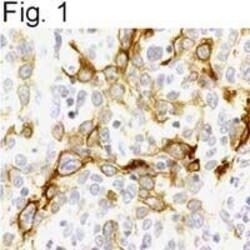
- Immunostaining of CXCR4 in human cervical carcinoma tissue sections.
Supportive validation
- Submitted by
- Invitrogen Antibodies (provider)
- Main image
- Experimental details
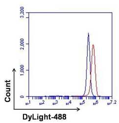
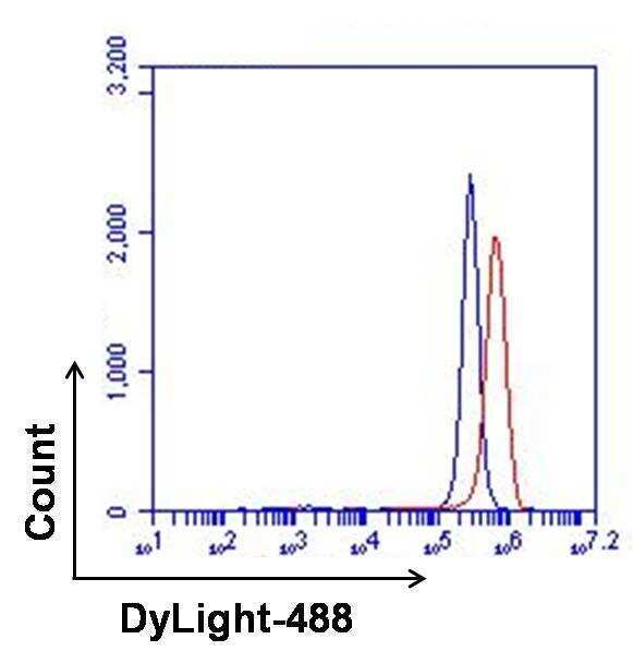
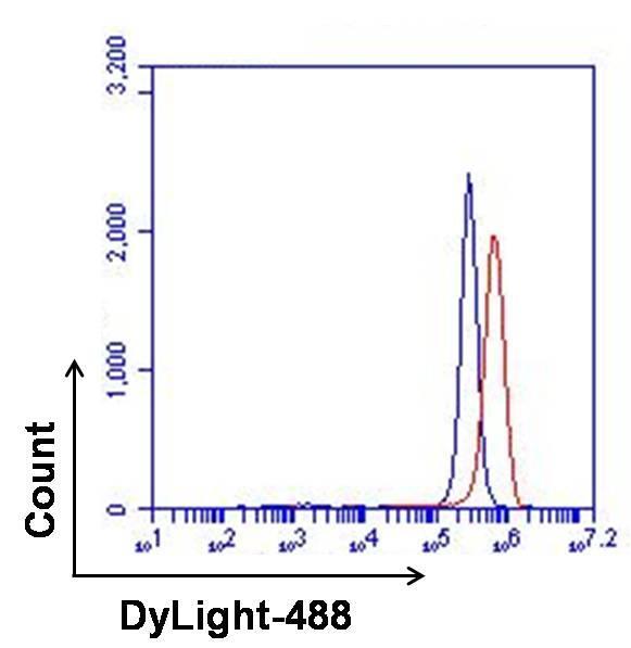
- Flow cytometry analysis of CXCR4 on Ramos cells. Cells were fixed, permeabilized, and then blocked with a 1:100 dilution of normal goat serum for at least 30 minutes on ice. Cells were stained with CXCR4 polyclonal antiserum (Product # PA3-305, red histogram) or with normal rabbit serum (blue histogram) at a dilution of 1:500. After incubation of the primary antibody for at least 1 hour on ice, the cells were stained with a DyLight 488-conjugated goat anti-rabbit IgG secondary antibody (Product # 35552) for at least 30 minutes on ice. A representative 10,000 cells were acquired for each sample.
- Submitted by
- Invitrogen Antibodies (provider)
- Main image
- Experimental details
- Flow cytometry analysis of CXCR4 on Ramos cells. Cells were fixed, permeabilized, and then blocked with a 1:100 dilution of normal goat serum for at least 30 minutes on ice. Cells were stained with CXCR4 polyclonal antiserum (Product # PA3-305, red histogram) or with normal rabbit serum (blue histogram) at a dilution of 1:500. After incubation of the primary antibody for at least 1 hour on ice, the cells were stained with a DyLight 488-conjugated goat anti-rabbit IgG secondary antibody (Product # 35552) for at least 30 minutes on ice. A representative 10,000 cells were acquired for each sample.
Supportive validation
- Submitted by
- Invitrogen Antibodies (provider)
- Main image

- Experimental details
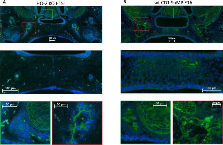
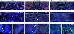
- FIGURE 7 CXCR4 expressing cells in the palatal osteogenic centers. (A) Upper panel: Fluorescent immunohistochemical-stained coronal palatal section for CXCR4 expression (green), representative for the wt E15 and HO-2 KO E15, e.g., HO-2 KO. Middle panel: Within the disintegrating MES almost no CXCR4 expressing cells were found. Green panel: Near the forming nasal septum clusters of strong CXCR4 expressing cells were found. Also within the cartilage of the forming nasal septum CXCR4 expressing cells were present. Red panel: The osteogenic centers in the lateral parts of the fusing palate demonstrated clusters of strong CXCR4 expressing cells. (B) Upper panel: Fluorescent immunohistochemical stained coronal palatal section for CXCR4 expression (green), representative for the wt CD1 E16 and wt CD1 SnMP E16 fetuses, e.g., wt CD1 SnMP E16. Middle panel: in the osteogenic centers in the central part of the fusing palate clusters of CXCR4-positive-stained mesenchymal cells were found. Green panel: Near the forming nasal septum clusters of strong CXCR4 expressing cells were found. Also within the cartilage of the forming nasal septum CXCR4 expressing cells were present. Red panel: The osteogenic centers in the lateral parts of the fusing palate demonstrated clusters of strong CXCR4 expressing cells.
- Submitted by
- Invitrogen Antibodies (provider)
- Main image
- Experimental details
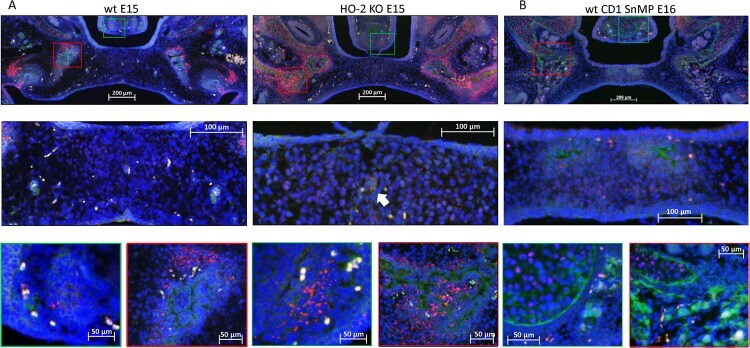
- FIGURE 8 Most CXCR4-positive cells in the palatal osteogenic centers are not positive for Sox9. (A) Upper panel: Fluorescent immunohistochemical double-stained coronal palatal section for Sox9 (red) with CXCR4 (green), representative for the wt E15 and HO-2 KO E15 fetuses. Middle panel: Near the disintegrating MES only a few Sox9-CXCR4 double-positive stained cells (white arrow) were found. Green panel: In the osteogenic centers near the forming nasal septum clusters of Sox9 expressing cells together with CXCR4 expressing cells. Red panel: The osteogenic centers in the lateral parts of the fusing palate demonstrated clusters of Sox9 expressing cells near CXCR4 expressing cells. (B) Upper panel: Fluorescent histochemical double-stained coronal palatal section for Sox9 (red) with CXCR4 (green), representative for the wt CD1 E16 and wt CD1 SnMP E16 fetuses, e.g., wt CD1 SnMP E16. Middle panel: In the osteogenic centers in the central part of the fusing palate, almost no Sox9-CXCR4 double-positive stained cells were found. Green panel: Near the forming nasal septum clusters of CXCR4 expressing cells were found near CXCR4 positive cells. Also in the cartilage of the nasal septum CXCR4 positive cells were found close to Sox9 positive cells. Red panel: The osteogenic centers in the lateral parts of the fusing palate demonstrated clusters of Sox9 expressing cells near CXCR4 expressing cells.
- Submitted by
- Invitrogen Antibodies (provider)
- Main image

- Experimental details
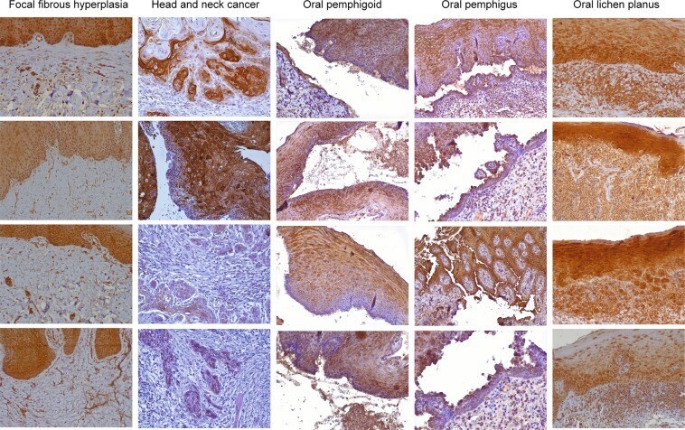
- Figure 3 CXCR4 is overexpressed in the connective tissue of oral lichen planus. Representative microphotographs of CXCR4 receptor immunohistochemical staining in oral tissues (40x objective). Positive staining is shown in brown. Compared to the controls, oral lichen planus lesions show high intensity and reactivity to CXCR4 in areas of active inflammatory infiltrate. All images (n = 99) can be reviewed at, 10.5281/zenodo.3352836.
- Submitted by
- Invitrogen Antibodies (provider)
- Main image
- Experimental details
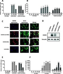
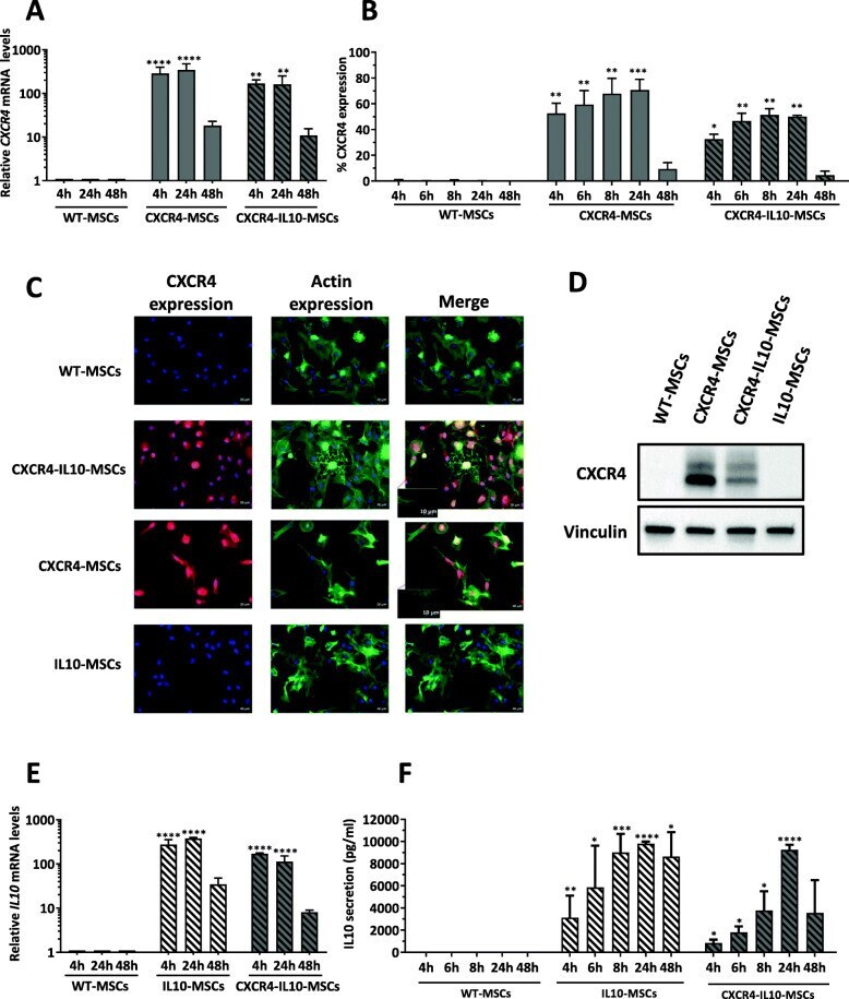
- Fig. 1 Enhanced expression of CXCR4 and IL10 in human Ad-MSCs transfected with specific mRNAs. a Analysis of CXCR4 mRNA levels at different times after transfection with CXCR4-mRNA and CXCR4-IL10-mRNA. b Flow cytometry analysis of CXCR4 in WT and mRNA-transfected Ad-MSCs at different times post-transfection. c Immunofluorescence staining of CXCR4 in WT and mRNA-transfected MSCs 4 h after transfection with the different mRNAs (bar = 20 mum). Inserts with higher magnifications (red frame) indicate that CXCR4-expressing MSCs resulted in the protrusion of long filopodia at the cell periphery. Bar = 10 mum. d CXCR4 expression analyzed by western blot 4 h after transfection with the different mRNAs using vinculin as a control. e Analysis of mRNA IL10 levels analyzed at different time points after transfection with IL10-mRNA and CXCR4-IL10-mRNAs. f IL10 secretion measured by ELISA in WT and mRNA-transfected Ad-MSCs at different time points after transfection. Bars in the plots represent mean +- SD from three independent experiments, and statistical differences were analyzed by Tukey's multiple comparison test. * p < 0.05, ** p < 0.01, *** p < 0.001, **** p < 0.0001; versus WT-MSCs
- Submitted by
- Invitrogen Antibodies (provider)
- Main image
- Experimental details
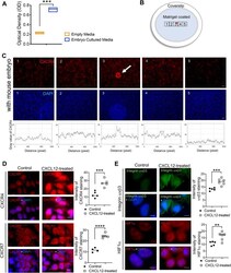
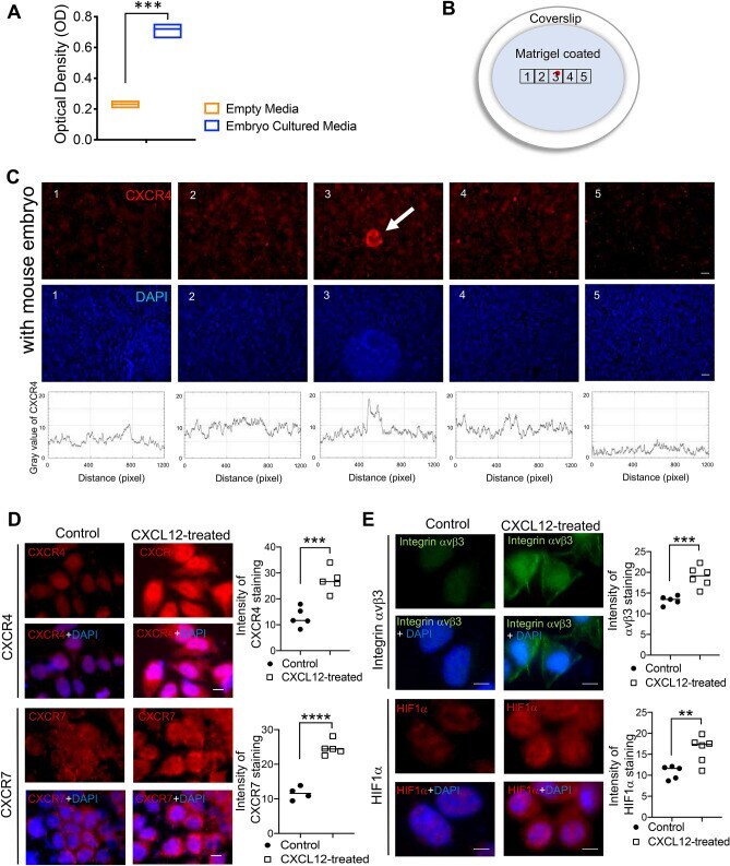
- Figure 1 Embryo-derived chemokine CXCL12 and its receptor endometrial CXCR4/CXCR7. ( A ) CXCL12 levels were quantified in embryo culture or empty medium by ELISA. Comparison groups are from 3 independent experiments and analyzed using the unpaired Student t test for parametric distributions including p-values (***< 0.001). ( B ) A schematic diagram of co-culture system of mouse embryo with Ishikawa cells on Matrigel-coated coverslip. ( C ) Immunofluorescence (IF) staining images (1-5) were obtained from 5 different regions (illustrated on a diagram of ( B:1-5 )) of coverslip and mouse embryo (white arrow) was attached in the region number 3 (illustrated on a diagram of ( B ), marked as red spot). Red: CXCR4, Blue: DAPI. Scale Bar; 50 mum. Intensity of CXCR4 in each image was profiled. Images were captured at a magnification of 10 x. IF staining of CXCR4, CXCR7 ( D ) and integrin vbeta3, HIF1 ( E ) in Ishikawa cells in response to CXCL12. Saline-treated cells were used for control. Scale Bar; 20 mum. Intensity of IF staining was quantified using Image J and displayed in graphs. Data are from 4 independent experiments, and analyzed using unpaired Student t test analysis including p-values (*< 0.05, **< 0.01, ***< 0.001, NS; not significant).
- Submitted by
- Invitrogen Antibodies (provider)
- Main image
- Experimental details
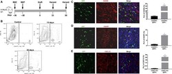
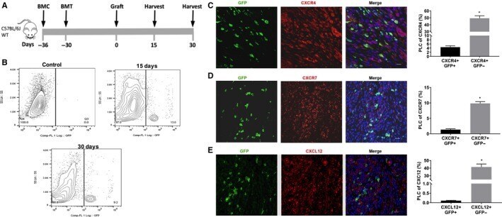
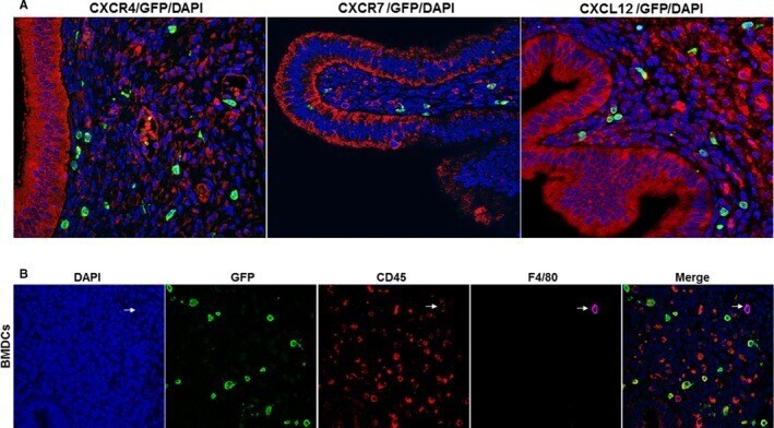
- Figure 1 A model of 5-FU submyeloablation to characterize the bone marrow cell trafficking into endometriotic lesions in mice. Schematic model: Treatment for bone marrow conditioning (BMC) started on day -36 before uterine fragment grafting and 6 days before bone marrow transplantation (BMT) (A). Ectopic lesions were harvested 15 and 30 days (each group n = 6) after grafting uterine fragments into the peritoneal. Representative images of flow cytometry analysis of ectopic lesion cells on day 15 and 30 after uterine fragment grafting demonstrating the percentage of GFP-positive cells in endometriosis. B, Control group (n = 6 mice), represented by GFP-negative endometriosis lesions, was used to set a gate. Fluorescence confocal microscopy analysis of mouse endometriosis sections (C, D and E). Representative images of IF and quantitative analyses of cells expressing CXCR4, CXCR7 or CXCL12 and GFP, as a per cent of total endometriosis cells. Murine lesions were stained with anti-GFP antibody (green) and co-stained with CXCR4 antibody (red) (C), with CXCR7 antibody (red) (D) and with CXCL12 antibody (red) (E). Nuclei were stained by DAPI and are shown in blue. BMC, Bone morrow conditioning; BMT, Bone morrow transplantation; PLC, percentage of labelled cells. Results shown as mean +- SE. * P < .05 vs vehicle. Scale bar: 20 mum
- Submitted by
- Invitrogen Antibodies (provider)
- Main image
- Experimental details
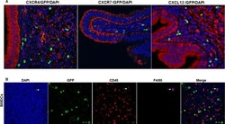
- Figure 2 Immunostaining of CXCR4, CXCR7 and CXCL12 and BMDCs. A, Representative immunofluorescence images of CXCR4, CXCR7 and CXCL12 staining in the epithelial cells of murine endometriosis. In our model, no GFP (bone marrow-derived cells) was identified in the epithelium. Scale bar: 10 mum. B, Engrafted BMDCs express CD45 and F4/80. BMDCs: Engrafted bone marrow-derived immune cells (BMDCs) were stained with DAPI or immunofluorescence for CD45 and F4/80. Engrafted BMDCs shows the presence of non-immune (CD45 negative) and immune cells (CD45 positive), some of which co-express F4/80, a marker of macrophages. Included is a cell that is a bone marrow-derived (GFP+), CD45+, F4/80+ macrophage (arrow) Scale bar: 10 mum
- Submitted by
- Invitrogen Antibodies (provider)
- Main image
- Experimental details
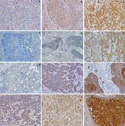
- Figure 1 Low/negative, moderate and high staining intensity for CCR4 (A,B,C), CCR5 (D,E,F), CCR7 (G,H,I) and CXCR4 (J,K,L) (20x objective).
