 Explore
 Validate
 Learn
LearnMMCP1I
antibody from Invitrogen Antibodies
Targeting: CCL2
GDCF-2, HC11, MCAF, MCP-1, MCP1, MGC9434, SCYA2, SMC-CF
 Western blot
 ELISA
ELISAAntibody data
- Antibody Data
- Antigen structure
- References [0]
- Comments [0]
- Validations
- Western blot [1]
- Immunocytochemistry [1]
Submit
Validation data
Reference
Comment
Report error
- Product number
- MMCP1I - Provider product page

- Provider
- Invitrogen Antibodies
- Product name
- MCP-1 Monoclonal Antibody (E10051)
- Antibody type
- Monoclonal
- Antigen
- Recombinant full-length protein
- Description
- MMCP1I targets MCP-1 in ELISA, and WB applications and shows reactivity with Human samples. The MMCP1I immunogen is recombinant human MCP-1. MMCP1I detects MCP-1 which has a predicted molecular weight of approximately 9 kDa. This product has been tested for endotoxins by limulus amoebocyte lysate (LAL) assay and contains an endotoxin concentration of less than or equal to 10 endotoxin units per milligram (EU/mg).
- Reactivity
- Human
- Host
- Mouse
- Isotype
- IgG
- Antibody clone number
- E10051
- Vial size
- 500 µg
- Concentration
- 1.0 mg/mL
- Storage
- -20°C
No comments: Submit comment
Supportive validation
- Submitted by
- Invitrogen Antibodies (provider)
- Main image
- Experimental details
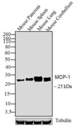
- Western blot analysis was performed on tissue extracts (30 µg lysate) of Mouse Pancreas (Lane 1), Mouse Spleen (Lane 2), Mouse Lung (Lane 3) and Mouse Cerebellum (Lane 4). The blots were probed with Anti-MCP-1 Mouse monoclonal Antibody (Product # MMCP1I, 1-2 µg/mL) and detected by chemiluminescence using Goat anti-Mouse IgG (H+L) Superclonal™ Secondary Antibody, HRP conjugate (Product # A28177, 0.4 µg/mL, 1:2500 dilution). A ~ 25 kDa band corresponding to MCP-1 was observed across cell lines. A shift in molecular weight is due to the glycosylation of MCP-1 protein.Known quantity of protein samples were electrophoresed using Novex® NuPAGE® 12 % Bis-Tris gel (Product # NP0342BOX), XCell SureLock™ Electrophoresis System (Product # EI0002) and Novex® Sharp Pre-Stained Protein Standard (Product # LC5800). Resolved proteins were then transferred onto a nitrocellulose membrane with iBlot® 2 Dry Blotting System (Product # IB21001). The membrane was probed with the relevant primary and secondary Antibody following blocking with 5 % skimmed milk. Chemiluminescent detection was performed using Pierce™ ECL Western Blotting Substrate (Product # 32106).

Supportive validation
- Submitted by
- Invitrogen Antibodies (provider)
- Main image
- Experimental details
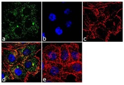
- Immunofluorescence analysis of MCP-1 was performed using 70% confluent log phase HCT 116 cells. The cells were fixed with 4% paraformaldehyde for 10 minutes, permeabilized with 0.1% Triton™ X-100 for 10 minutes, and blocked with 2% BSA for 1 hour at room temperature. The cells were labeled with MCP-1 (E10051) Mouse Monoclonal Antibody (MMCP1I) at 2 µg/mL in 0.1% BSA and incubated for 3 hours at room temperature and then labeled with Goat anti-Mouse IgG (H+L) Superclonal™ Secondary Antibody, Alexa Fluor® 488 conjugate (Product # A28175) a dilution of 1:2000 for 45 minutes at room temperature (Panel a: green). Nuclei (Panel b: blue) were stained with SlowFade® Gold Antifade Mountant with DAPI (Product # S36938). F-actin (Panel c: red) was stained with Alexa Fluor® 555 Rhodamine Phalloidin (Product # R415, 1:300). Panel d represents the merged image showing punctate cytoplasmic localization. Panel e shows the no primary antibody control. The images were captured at 60X magnification.
