Explore
 Validate
 Learn
Learn12-7096-82
antibody from Invitrogen Antibodies
Targeting: CCL2
GDCF-2, HC11, MCAF, MCP-1, MCP1, MGC9434, SCYA2, SMC-CF
 Flow cytometry
Antibody data
- Antibody Data
- Antigen structure
- References [5]
- Comments [0]
- Validations
- Flow cytometry [1]
- Other assay [1]
Submit
Validation data
Reference
Comment
Report error
- Product number
- 12-7096-82 - Provider product page

- Provider
- Invitrogen Antibodies
- Product name
- CCL2 (MCP-1) Monoclonal Antibody (2H5), PE, eBioscience™
- Antibody type
- Monoclonal
- Antigen
- Other
- Description
- Description: The 2H5 monoclonal antibody reacts with mouse, rat, and human monocyte chemoattractant protein-1 (MCP-1), also known as CCL2 and MCAF. Applications Reported:The 2H5 antibody has been reported for use in ELISA, intracellular staining for flow cytometric analysis, and neutralization. (Please use Functional Grade purified 2H5 (Product # 16-7096-81) in functional assays). Applications Tested: This 2H5 antibody is tested by flow cytometric analysis of fixed permabilized LPS-stimulated mouse thioglycolate-elicited peritoneal exudate cells. Has been tested by flow cytometric analysis of fixed permabilized LPS-stimulated mouse thioglycolate-elicited peritoneal exudate cells. This can be used at less than or equal to 0.25 µg per test. A test is defined as the amount (µg) of antibody that will stain a cell sample in a final volume of 100 µL. Cell number should be determined empirically but can range from 10^5 to 10^8 cells/test. It is recommended that the antibody be carefully titrated for optimal performance in the assay of interest. Excitation: 488-561 nm; Emission: 578 nm; Laser: Blue Laser, Green Laser, Yellow-Green Laser. Filtration: 0.2 µm post-manufacturing filtered.
- Reactivity
- Human, Mouse, Rat
- Conjugate
- Yellow dye
- Isotype
- IgG
- Antibody clone number
- 2H5
- Vial size
- 100 µg
- Concentration
- 0.2 mg/mL
- Storage
- 4° C, store in dark, DO NOT FREEZE!
Submitted references Lysosomal acid lipase, CSF1R, and PD-L1 determine functions of CD11c+ myeloid-derived suppressor cells.
Conditioned medium from umbilical cord mesenchymal stem cells induces migration and angiogenesis.
Mechanisms underlying renoprotection during renin-angiotensin system blockade.
Mechanisms underlying renoprotection during renin-angiotensin system blockade.
Macrophage inflammatory protein-2 and KC induce chemokine production by mouse astrocytes.
Zhao T, Liu S, Ding X, Johnson EM, Hanna NH, Singh K, Sen CK, Wan J, Du H, Yan C
JCI insight 2022 Sep 8;7(17)
JCI insight 2022 Sep 8;7(17)
Conditioned medium from umbilical cord mesenchymal stem cells induces migration and angiogenesis.
Shen C, Lie P, Miao T, Yu M, Lu Q, Feng T, Li J, Zu T, Liu X, Li H
Molecular medicine reports 2015 Jul;12(1):20-30
Molecular medicine reports 2015 Jul;12(1):20-30
Mechanisms underlying renoprotection during renin-angiotensin system blockade.
Taal MW, Chertow GM, Rennke HG, Gurnani A, Jiang T, Shahsafaei A, Troy JL, Brenner BM, Mackenzie HS
American journal of physiology. Renal physiology 2001 Feb;280(2):F343-55
American journal of physiology. Renal physiology 2001 Feb;280(2):F343-55
Macrophage inflammatory protein-2 and KC induce chemokine production by mouse astrocytes.
Luo Y, Fischer FR, Hancock WW, Dorf ME
Journal of immunology (Baltimore, Md. : 1950) 2000 Oct 1;165(7):4015-23
Journal of immunology (Baltimore, Md. : 1950) 2000 Oct 1;165(7):4015-23
No comments: Submit comment
Supportive validation
- Submitted by
- Invitrogen Antibodies (provider)
- Main image
- Experimental details
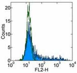
- Staining of 24-hour LPS-stimulated normal human peripheral blood cells with 0.25 µg of Armenian Hamster IgG Isotype Control PE (Product # 12-4888-81) (open histogram) or 0.25 µg of Anti-CCL2 (MCP-1) PE (filled histogram). Cells in the monocyte gate were used for analysis.
- Conjugate
- Yellow dye

Supportive validation
- Submitted by
- Invitrogen Antibodies (provider)
- Main image
- Experimental details
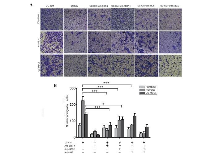
- Figure 5 Migration of fibroblasts, HUVECs and UC-MSCs in response to UC-CM. (A) A total of 5x10 4 cells were collected and allowed to migrate. Lane 1, UC-CM; lane 2, DMEM; lanes 3-6, in the presence or absence of anti-SDF-1 (20 mu g/ml), anti-MCP-1 (20 mu g/ml) or anti-HGF (20 mu g/ml), respectively. Results are from a representative experiment and are expressed as the mean number of migrated cells in three random fields, scale bar=200 mu m. Cells that crossed the matrigel membrane were stained with crystal violet (magnification, x40). (B) Graphical presentation of the quantified data, presented as the number of migrated cells and expressed as the mean +- standard error of the mean. HUVECs, human umbilical vein endothelial cells; UC-MSCs, umbilical cord mesenchymal stem cells; UC-CM, UC-MSCs conditioned medium; DMEM, Dulbecco's modified Eagle's medium; SDF-1, stromal cell-derived factor 1; MCP-1, monocyte chemotactic protein 1; HGF, hepatocyte growth factor.
- Conjugate
- Yellow dye