Explore
 Validate
 Learn
Learn Immunocytochemistry
Antibody data
- Antibody Data
- Antigen structure
- References [1]
- Comments [0]
- Validations
- Immunocytochemistry [1]
- Immunohistochemistry [1]
Submit
Validation data
Reference
Comment
Report error
- Product number
- HPA001583 - Provider product page

- Provider
- Atlas Antibodies
- Proper citation
- Atlas Antibodies Cat#HPA001583, RRID:AB_1079641
- Product name
- Anti-ATP2B3
- Antibody type
- Polyclonal
- Description
- Polyclonal Antibody against Human ATP2B3, Gene description: ATPase, Ca++ transporting, plasma membrane 3, Alternative Gene Names: CFAP39, CLA2, PMCA3, SCAX1, Validated applications: ICC, IHC, Uniprot ID: Q16720, Storage: Store at +4°C for short term storage. Long time storage is recommended at -20°C.
- Reactivity
- Human
- Host
- Rabbit
- Conjugate
- Unconjugated
- Isotype
- IgG
- Vial size
- 100 µl
- Concentration
- 0.1 mg/ml
- Storage
- Store at +4°C for short term storage. Long time storage is recommended at -20°C.
- Handling
- The antibody solution should be gently mixed before use.
Submitted references Somatic mutations in ATP1A1 and ATP2B3 lead to aldosterone-producing adenomas and secondary hypertension
Beuschlein F, Boulkroun S, Osswald A, Wieland T, Nielsen H, Lichtenauer U, Penton D, Schack V, Amar L, Fischer E, Walther A, Tauber P, Schwarzmayr T, Diener S, Graf E, Allolio B, Samson-Couterie B, Benecke A, Quinkler M, Fallo F, Plouin P, Mantero F, Meitinger T, Mulatero P, Jeunemaitre X, Warth R, Vilsen B, Zennaro M, Strom T, Reincke M
Nature Genetics 2013;45(4):440-444
Nature Genetics 2013;45(4):440-444
No comments: Submit comment
Supportive validation
- Submitted by
- Atlas Antibodies (provider)
- Main image
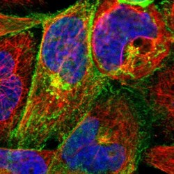
- Experimental details
- Immunofluorescent staining of human cell line A-431 shows localization to plasma membrane & the Golgi apparatus.
- Sample type
- Human

Supportive validation
- Submitted by
- Atlas Antibodies (provider)
- Enhanced method
- Orthogonal validation
- Main image
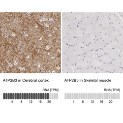
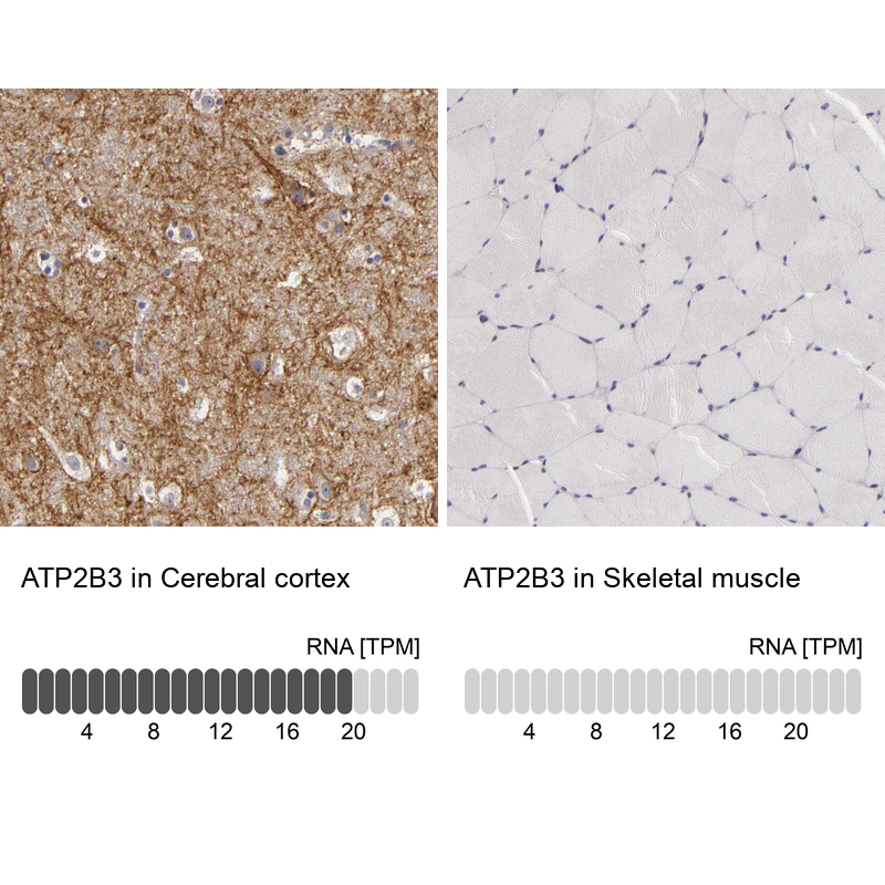
- Experimental details
- Immunohistochemistry analysis in human cerebral cortex and skeletal muscle tissues using HPA001583 antibody. Corresponding ATP2B3 RNA-seq data are presented for the same tissues.
- Sample type
- Human
- Protocol
- Protocol