Explore
 Validate
 Learn
Learn Western blot
Antibody data
- Antibody Data
- Antigen structure
- References [2]
- Comments [0]
- Validations
- Western blot [1]
- Immunohistochemistry [2]
Submit
Validation data
Reference
Comment
Report error
- Product number
- MAB41051 - Provider product page

- Provider
- R&D Systems
- Product name
- Human/Mouse Patched 1/PTCH (First Extracellular Loop) Antibody
- Antibody type
- Monoclonal
- Description
- Protein A or G purified from hybridoma culture supernatant. Detects mouse Patched 1/PTCH (First Extracellular Loop) in direct ELISAs and Western blots. In direct ELISAs and Western blots, 15% cross-reactivity with recombinant human Patched 2 is observed.
- Reactivity
- Human, Mouse
- Host
- Rat
- Conjugate
- Unconjugated
- Antigen sequence
Q61115- Isotype
- IgG
- Antibody clone number
- 413220
- Vial size
- 100 ug
- Concentration
- LYOPH
- Storage
- Use a manual defrost freezer and avoid repeated freeze-thaw cycles. 12 months from date of receipt, -20 to -70 °C as supplied. 1 month, 2 to 8 °C under sterile conditions after reconstitution. 6 months, -20 to -70 °C under sterile conditions after reconstitution.
Submitted references Wnt Signaling Separates the Progenitor and Endocrine Compartments during Pancreas Development.
Defective Wnt-dependent cerebellar midline fusion in a mouse model of Joubert syndrome.
Sharon N, Vanderhooft J, Straubhaar J, Mueller J, Chawla R, Zhou Q, Engquist EN, Trapnell C, Gifford DK, Melton DA
Cell reports 2019 May 21;27(8):2281-2291.e5
Cell reports 2019 May 21;27(8):2281-2291.e5
Defective Wnt-dependent cerebellar midline fusion in a mouse model of Joubert syndrome.
Lancaster MA, Gopal DJ, Kim J, Saleem SN, Silhavy JL, Louie CM, Thacker BE, Williams Y, Zaki MS, Gleeson JG
Nature medicine 2011 Jun;17(6):726-31
Nature medicine 2011 Jun;17(6):726-31
No comments: Submit comment
Supportive validation
- Submitted by
- R&D Systems (provider)
- Main image
- Experimental details
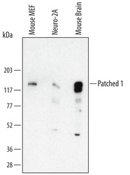
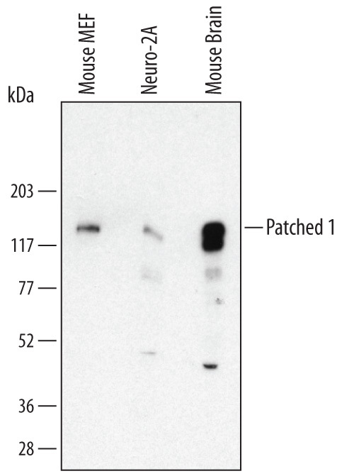
- Detection of Mouse Patched 1/PTCH by Western Blot. Western blot shows lysates of MEF mouse embryonic feeder cells, Neuro-2A mouse neuroblastoma cell line, and mouse brain tissue. PVDF Membrane was probed with 1 µg/mL of Rat Anti-Human/Mouse Patched 1/PTCH (First Extracellular Loop) Monoclonal Antibody (Catalog # MAB41051) followed by HRP-conjugated Anti-Rat IgG Secondary Antibody (Catalog # HAF005). A specific band was detected for Patched 1/PTCH at approximately 160 kDa (as indicated). This experiment was conducted under reducing conditions and using Immunoblot Buffer Group 1.
Supportive validation
- Submitted by
- R&D Systems (provider)
- Main image
- Experimental details
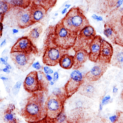
- Patched 1/PTCH in Human Esophageal Squamous Cell Carcinoma. Patched 1/PTCH was detected in immersion fixed paraffin-embedded sections of human esophogeal squamous cell carcinoma tissue using Rat Anti-Human/Mouse Patched 1/PTCH (First Extracellular Loop) Monoclonal Antibody (Catalog # MAB41051) at 15 µg/mL overnight at 4 °C. Tissue was stained using the Anti-Mouse HRP-DAB Cell & Tissue Staining Kit (brown; Catalog # CTS002) and counterstained with hematoxylin (blue). Specific staining was localized to plasma membranes. View our protocol for Chromogenic IHC Staining of Paraffin-embedded Tissue Sections.

- Submitted by
- R&D Systems (provider)
- Main image
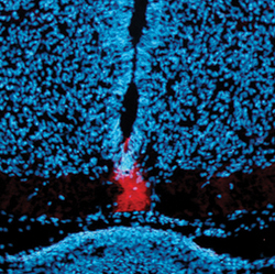
- Experimental details
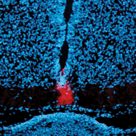
- Patched in Mouse Spinal Cord. Patched was detected in immersion fixed frozen sections of mouse spinal cord using 10 µg/mL Rat Anti-Human/Mouse Patched 1/PTCH (First Extracellular Loop) Monoclonal Antibody (Catalog # MAB41051) overnight at 4 °C. Tissue was stained (red) and counterstained with DAPI (blue). View our protocol for Fluorescent IHC Staining of Frozen Tissue Sections.