Explore
 Validate
 Learn
Learn Immunocytochemistry
Antibody data
- Antibody Data
- Antigen structure
- References [0]
- Comments [0]
- Validations
- Immunocytochemistry [2]
- Immunohistochemistry [3]
Submit
Validation data
Reference
Comment
Report error
- Product number
- PA5-54060 - Provider product page

- Provider
- Invitrogen Antibodies
- Product name
- PSIP1 Polyclonal Antibody
- Antibody type
- Polyclonal
- Antigen
- Recombinant full-length protein
- Description
- Immunogen sequence: KQVETEEAGV VTTATASVNL KVSPKRGRPA ATEVKIPKPR GRPKMVKQPC PSESDIITEE DKSKKKGQEE KQPKKQPKKD EEGQ
- Concentration
- 0.4 mg/mL
No comments: Submit comment
Supportive validation
- Submitted by
- Invitrogen Antibodies (provider)
- Main image

- Experimental details
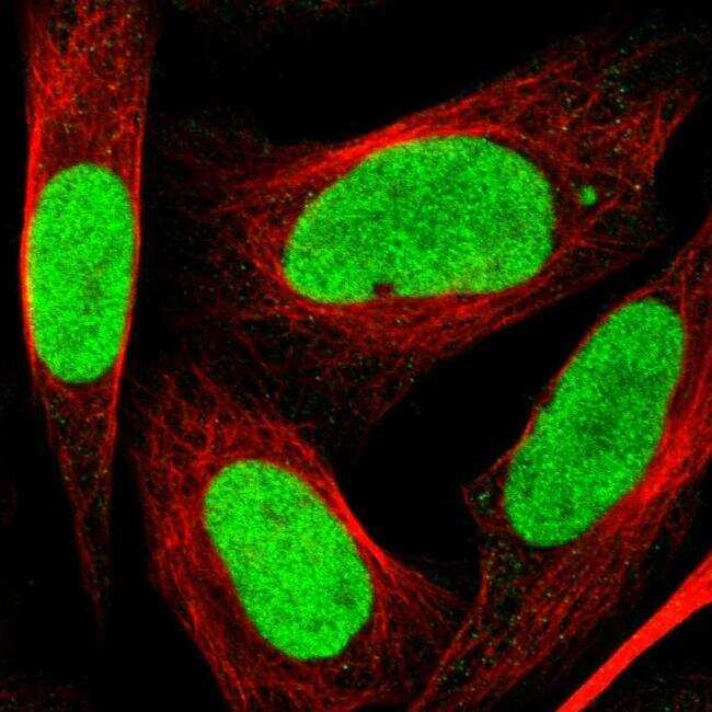
- Immunofluorescent staining of PSIP1 in human cell line U-2 OS shows positivity in nucleus but excluded from the nucleoli. Samples were probed using a PSIP1 Polyclonal Antibody (Product # PA5-54060).
- Submitted by
- Invitrogen Antibodies (provider)
- Main image

- Experimental details
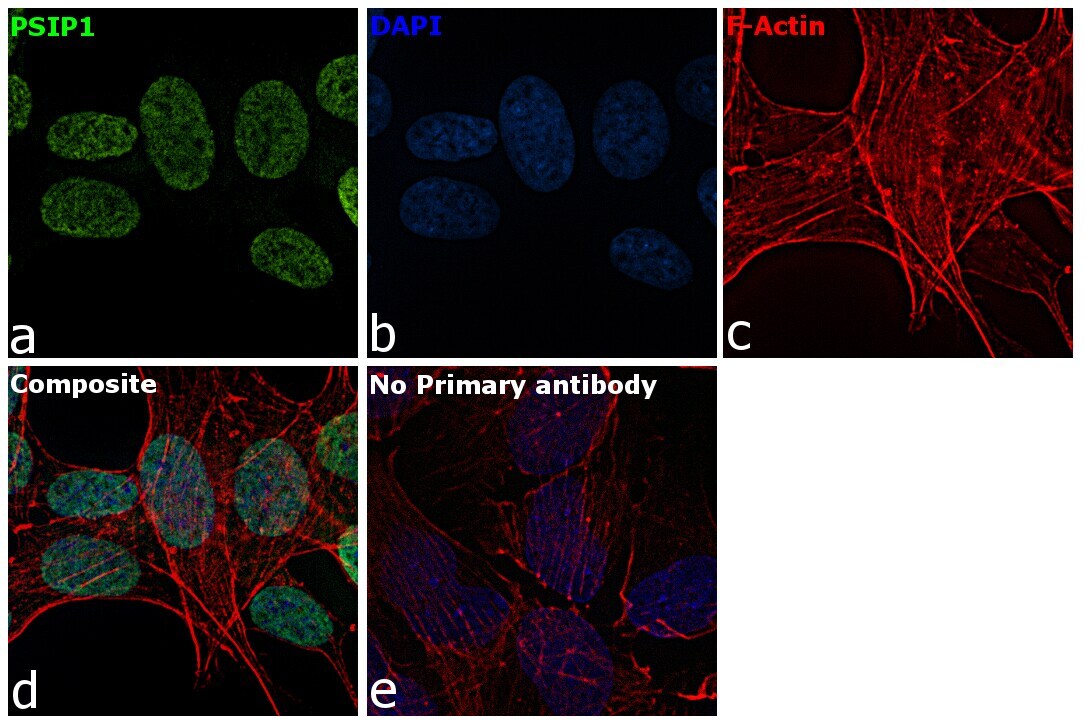
- Immunofluorescence analysis of PSIP1 was performed using SH-SY5Y cells. The cells were fixed with 4% paraformaldehyde for 10 minutes, permeabilized with 0.1% Triton™ X-100 for 10 minutes, and blocked with 2% BSA for 1 hour at room temperature. The cells were labeled with PSIP1 Rabbit Polyclonal Antibody (Product # PA5-54060) at 4 µg/mL in 0.1% BSA and incubated overnight at 4 degree and then labeled with Goat anti-Rabbit IgG (H+L) Superclonal™ Secondary Antibody, Alexa Fluor® 488 conjugate (Product # A27034) at a dilution of 1:2000 for 45 minutes at room temperature (Panel a: green). Nuclei (Panel b: blue) were stained with SlowFade® Gold Antifade Mountant with DAPI (Product # S36938). F-actin (Panel c: red) was stained with Rhodamine Phalloidin (Product # R415, 1:300). Panel d represents the merged image showing nuclear localization. Panel e represents control cells with no primary antibody to assess background. The images were captured at 60X magnification.
Supportive validation
- Submitted by
- Invitrogen Antibodies (provider)
- Main image
- Experimental details
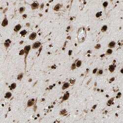
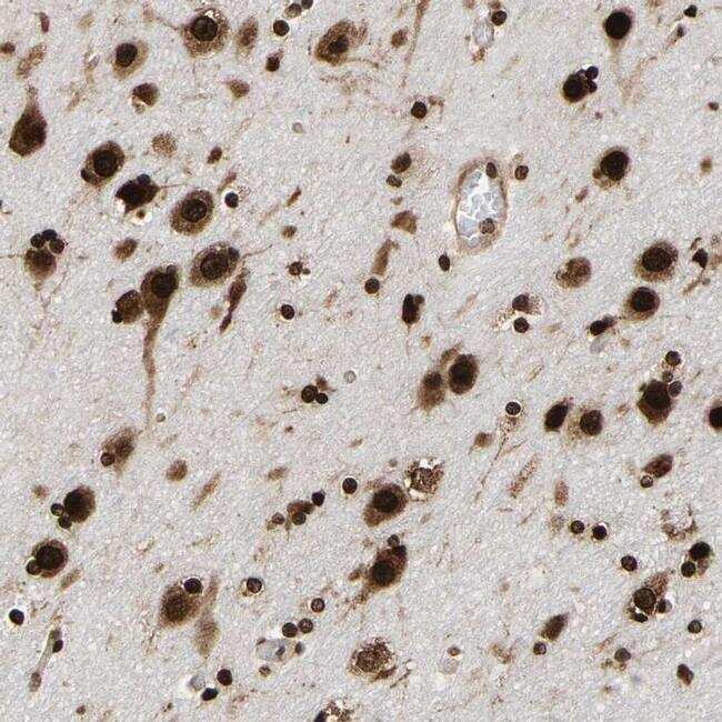
- Immunohistochemical staining of PSIP1 in human cerebral cortex tissue shows strong nuclear as well as cytoplasmic positivity in neuronal and glial cells. Samples were probed using a PSIP1 Polyclonal Antibody (Product # PA5-54060).
- Submitted by
- Invitrogen Antibodies (provider)
- Main image

- Experimental details
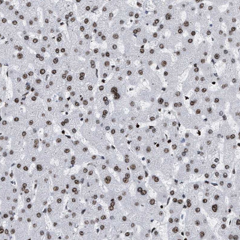
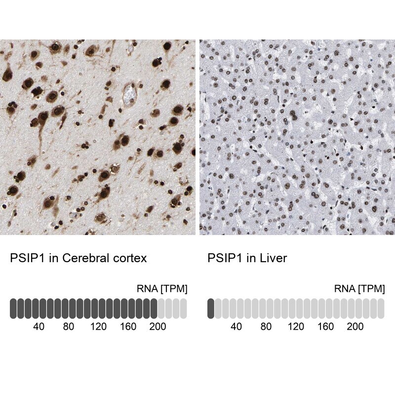
- Immunohistochemical staining of PSIP1 in human cerebral cortex and liver tissues using PSIP1 Polyclonal Antibody (Product # PA5-54060). Corresponding PSIP1 RNA-seq data are presented for the same tissues.
- Submitted by
- Invitrogen Antibodies (provider)
- Main image

- Experimental details
- Immunohistochemical staining of PSIP1 in human liver using PSIP1 Polyclonal Antibody (Product # PA5-54060) shows low expression as expected.