 Explore
 Validate
 Learn
Learn Western blot
 Immunocytochemistry
Antibody data
- Antibody Data
- Antigen structure
- References [10]
- Comments [0]
- Validations
- Immunocytochemistry [5]
- Immunoprecipitation [1]
- Immunohistochemistry [3]
- Other assay [6]
Submit
Validation data
Reference
Comment
Report error
- Product number
- PA5-27137 - Provider product page

- Provider
- Invitrogen Antibodies
- Product name
- ACSL4 Polyclonal Antibody
- Antibody type
- Polyclonal
- Antigen
- Synthetic peptide
- Description
- Recommended positive controls: MCF-7, MDA-MB-231, NIH-3T3, JC, BCL-1. Predicted reactivity: Mouse (100%), Rat (100%), Pig (100%), Rhesus Monkey (100%). Store product as a concentrated solution. Centrifuge briefly prior to opening the vial.
- Reactivity
- Human, Mouse, Rat
- Host
- Rabbit
- Isotype
- IgG
- Vial size
- 100 μL
- Concentration
- 1.37 mg/mL
- Storage
- Store at 4°C short term. For long term storage, store at -20°C, avoiding freeze/thaw cycles.
Submitted references Dipeptidase-1 governs renal inflammation during ischemia reperfusion injury.
Embryonal erythropoiesis and aging exploit ferroptosis.
Dual-process brain mitochondria isolation preserves function and clarifies protein composition.
Tumor suppressor p53 promotes ferroptosis in oxidative stress conditions independent of modulation of ferroptosis by p21, CDKs, RB, and E2F.
GSAP regulates lipid homeostasis and mitochondrial function associated with Alzheimer's disease.
Diet and Exercise Training Influence Skeletal Muscle Long-Chain acyl-CoA Synthetases.
Tissue-Specific Ablation of ACSL4 Results in Disturbed Steroidogenesis.
Control of antiviral innate immune response by protein geranylgeranylation.
BMP4 Upregulation Is Associated with Acquired Drug Resistance and Fatty Acid Metabolism in EGFR-Mutant Non-Small-Cell Lung Cancer Cells.
The endogenous subcellular localisations of the long chain fatty acid-activating enzymes ACSL3 and ACSL4 in sarcoma and breast cancer cells.
Lau A, Rahn JJ, Chappellaz M, Chung H, Benediktsson H, Bihan D, von Mässenhausen A, Linkermann A, Jenne CN, Robbins SM, Senger DL, Lewis IA, Chun J, Muruve DA
Science advances 2022 Feb 4;8(5):eabm0142
Science advances 2022 Feb 4;8(5):eabm0142
Embryonal erythropoiesis and aging exploit ferroptosis.
Zheng H, Jiang L, Tsuduki T, Conrad M, Toyokuni S
Redox biology 2021 Oct 30;48:102175
Redox biology 2021 Oct 30;48:102175
Dual-process brain mitochondria isolation preserves function and clarifies protein composition.
Noterman MF, Chaubey K, Lin-Rahardja K, Rajadhyaksha AM, Pieper AA, Taylor EB
Proceedings of the National Academy of Sciences of the United States of America 2021 Mar 16;118(11)
Proceedings of the National Academy of Sciences of the United States of America 2021 Mar 16;118(11)
Tumor suppressor p53 promotes ferroptosis in oxidative stress conditions independent of modulation of ferroptosis by p21, CDKs, RB, and E2F.
Kuganesan N, Dlamini S, Tillekeratne LMV, Taylor WR
The Journal of biological chemistry 2021 Dec;297(6):101365
The Journal of biological chemistry 2021 Dec;297(6):101365
GSAP regulates lipid homeostasis and mitochondrial function associated with Alzheimer's disease.
Xu P, Chang JC, Zhou X, Wang W, Bamkole M, Wong E, Bettayeb K, Jiang LL, Huang T, Luo W, Xu H, Nairn AC, Flajolet M, Ip NY, Li YM, Greengard P
The Journal of experimental medicine 2021 Aug 2;218(8)
The Journal of experimental medicine 2021 Aug 2;218(8)
Diet and Exercise Training Influence Skeletal Muscle Long-Chain acyl-CoA Synthetases.
Stierwalt HD, Ehrlicher SE, Robinson MM, Newsom SA
Medicine and science in sports and exercise 2020 Mar;52(3):569-576
Medicine and science in sports and exercise 2020 Mar;52(3):569-576
Tissue-Specific Ablation of ACSL4 Results in Disturbed Steroidogenesis.
Wang W, Hao X, Han L, Yan Z, Shen WJ, Dong D, Hasbargen K, Bittner S, Cortez Y, Greenberg AS, Azhar S, Kraemer FB
Endocrinology 2019 Nov 1;160(11):2517-2528
Endocrinology 2019 Nov 1;160(11):2517-2528
Control of antiviral innate immune response by protein geranylgeranylation.
Yang S, Harding AT, Sweeney C, Miao D, Swan G, Zhou C, Jiang Z, Fitzgerald KA, Hammer G, Bergo MO, Kroh HK, Lacy DB, Sun C, Glogauer M, Que LG, Heaton NS, Wang D
Science advances 2019 May;5(5):eaav7999
Science advances 2019 May;5(5):eaav7999
BMP4 Upregulation Is Associated with Acquired Drug Resistance and Fatty Acid Metabolism in EGFR-Mutant Non-Small-Cell Lung Cancer Cells.
Bach DH, Luu TT, Kim D, An YJ, Park S, Park HJ, Lee SK
Molecular therapy. Nucleic acids 2018 Sep 7;12:817-828
Molecular therapy. Nucleic acids 2018 Sep 7;12:817-828
The endogenous subcellular localisations of the long chain fatty acid-activating enzymes ACSL3 and ACSL4 in sarcoma and breast cancer cells.
Radif Y, Ndiaye H, Kalantzi V, Jacobs R, Hall A, Minogue S, Waugh MG
Molecular and cellular biochemistry 2018 Nov;448(1-2):275-286
Molecular and cellular biochemistry 2018 Nov;448(1-2):275-286
No comments: Submit comment
Supportive validation
- Submitted by
- Invitrogen Antibodies (provider)
- Main image
- Experimental details
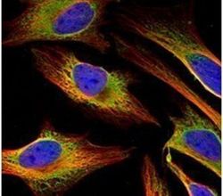
- Immunofluorescent analysis of FACL4 in methanol-fixed HeLa cells using a FACL4 polyclonal antibody (Product # PA5-27137) (Green) at a 1:500 dilution. Alpha-tubulin filaments were labeled with Product # PA5-29281 (Red) at a 1:2000.
- Submitted by
- Invitrogen Antibodies (provider)
- Main image
- Experimental details
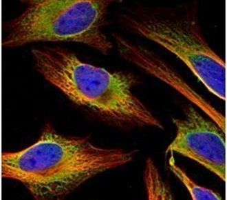
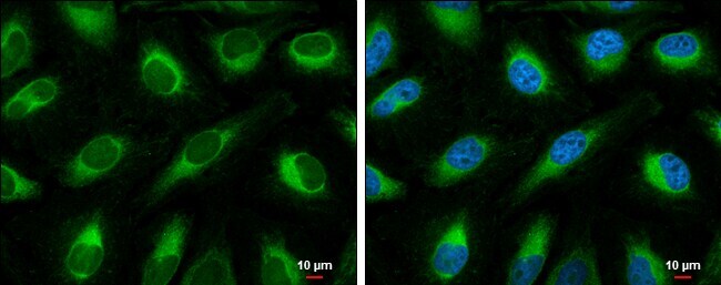
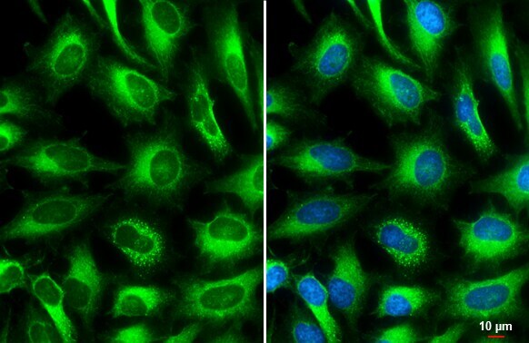
- Immunocytochemistry-Immunofluorescence analysis of ACSL4 was performed in HeLa cells fixed in 4% paraformaldehyde at RT for 15 min. Green: ACSL4 Polyclonal Antibody (Product # PA5-27137) diluted at 1:500. Blue: Hoechst 33342 staining. Scale bar = 10 µm.
- Submitted by
- Invitrogen Antibodies (provider)
- Main image

- Experimental details
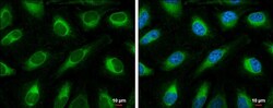
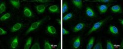
- ACSL4 Polyclonal Antibody detects FACL4 protein at cytoplasm by immunofluorescent analysis. Sample: HeLa cells were fixed in 4% paraformaldehyde at RT for 15 min. Green: FACL4 stained by ACSL4 Polyclonal Antibody (Product # PA5-27137) diluted at 1:500. Blue: Fluoroshield with DAPI . Scale bar= 10 µm.
- Submitted by
- Invitrogen Antibodies (provider)
- Main image
- Experimental details
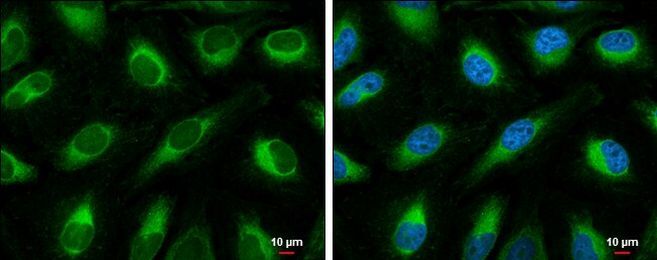
- Immunocytochemistry-Immunofluorescence analysis of ACSL4 was performed in HeLa cells fixed in 4% paraformaldehyde at RT for 15 min. Green: ACSL4 Polyclonal Antibody (Product # PA5-27137) diluted at 1:500. Blue: Hoechst 33342 staining. Scale bar = 10 µm.
- Submitted by
- Invitrogen Antibodies (provider)
- Main image
- Experimental details
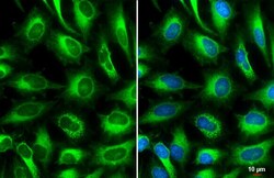
- ACSL4 Polyclonal Antibody detects FACL4 protein at cytoplasm by immunofluorescent analysis. Sample: HeLa cells were fixed in 4% paraformaldehyde at RT for 15 min. Green: FACL4 stained by ACSL4 Polyclonal Antibody (Product # PA5-27137) diluted at 1:500. Blue: Fluoroshield with DAPI . Scale bar= 10 µm.

Supportive validation
- Submitted by
- Invitrogen Antibodies (provider)
- Main image
- Experimental details
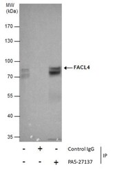
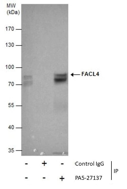
- Immunoprecipitation of FACL4 was performed in HeLa whole cell extracts using 5 µg of ACSL4 Polyclonal Antibody (Product # PA5-27137). Samples were transferred to a membrane and probed with ACSL4 Polyclonal Antibody as a primary antibody and an HRP-conjugated HRP-conjugated anti rabbit IgG was used as a secondary antibody.

Supportive validation
- Submitted by
- Invitrogen Antibodies (provider)
- Main image

- Experimental details
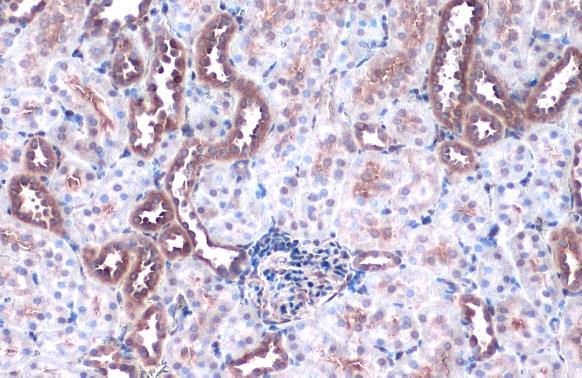
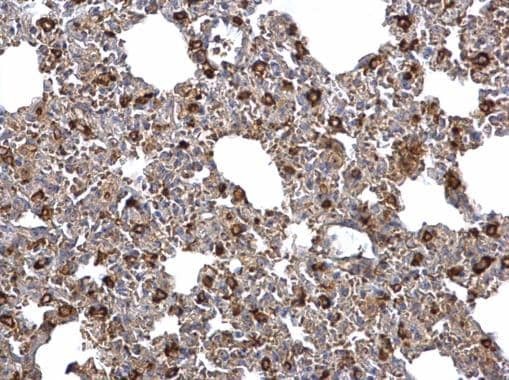
- ACSL4 Polyclonal Antibody detects FACL4 protein at cell membrane and cytoplasm by immunohistochemical analysis. Sample: Paraffin-embedded mouse kidney. FACL4 stained by ACSL4 Polyclonal Antibody (Product # PA5-27137) diluted at 1:500. Antigen Retrieval: Citrate buffer, pH 6.0, 15 min.
- Submitted by
- Invitrogen Antibodies (provider)
- Main image
- Experimental details
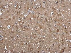
- ACSL4 Polyclonal Antibody detects FACL4 protein at cytosol on rat middle brain by immunohistochemical analysis. Sample: Paraffin-embedded rat middle brain. ACSL4 Polyclonal Antibody (Product # PA5-27137) dilution: 1:500. Antigen Retrieval: EDTA based buffer, pH 8.0, 15 min.
- Submitted by
- Invitrogen Antibodies (provider)
- Main image

- Experimental details
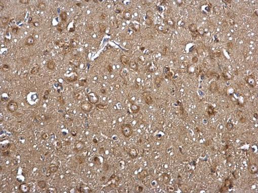
- ACSL4 Polyclonal Antibody detects FACL4 protein at cytosol on mouse lymph node by immunohistochemical analysis. Sample: Paraffin-embedded mouse lymph node. ACSL4 Polyclonal Antibody (Product # PA5-27137) dilution: 1:500. Antigen Retrieval: EDTA based buffer, pH 8.0, 15 min.
Supportive validation
- Submitted by
- Invitrogen Antibodies (provider)
- Main image
- Experimental details
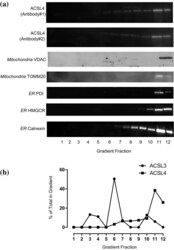
- Fig. 5 Equilibrium distributions of organelle marker proteins and ACSL4 in MCF-7 density gradient fractions. a Equal volume samples from each MCF-7 sucrose gradient fraction were subjected to SDS-PAGE separation and immunoblotted for ACSL4 using two different commercially available antibodies; antibody#1 was supplied by GeneTex and antibody#2 from Invitrogen. The gradient distributions of a panel of organelle marker proteins was similarly determined by immunoblotting. Data presented are representitive of experiments repeated 2-3 times with similar results. b The normalised distributions of anti-ACSL3 and anti-ACSL4 immunoreactivties in the gradient fractions. Western blotting signals were quantified using imageJ software. Data are representitive of experiments repeated 3-4 times with similar results

- Submitted by
- Invitrogen Antibodies (provider)
- Main image
- Experimental details
- Figure 5 BMP4 Knockdown Influences Metabolism and p53 (A) Heatmap showing and comparing top enriched terms. Enrichment test based on the gene ontology (GO, http://geneontology.org/ ) database was conducted using the significant gene list. Significant enrichments are displayed in blue (p value = 0.0001). (B) Heatmap representing changes in expression of top upregulated and downregulated genes in PC9-Gef cells transfected with control or BMP4 #2 siRNA. (C) PC9-Gef and H1993-Gef cells were transfected with control or BMP4 siRNA for 48 hr, then cell lysates were subjected to real-time PCR (top panels) or immunoblotting (bottom panels). (D) PC9-Gef and H1993-Gef cells were transfected with control or ACSL4 siRNA for 48 hr, then cell lysates were subjected to real-time PCR (top panels) or immunoblotting (bottom panels). (E) Schematic diagram illustrating the proposed BMP4 pathway modulating energy metabolism through ACSL4 and triglycerides. (F) PC9-Gef and H1993-Gef cells were transfected with control and BMP4 #2 siRNA for 48 hr, and then cell lysates were further processed for metabolic analyses as described in the Materials and Methods . (G) PC9-Gef cells were transfected with control or siBMP4 #2 siRNA for 48 hr, and cell lysates were subjected to the phosphor-kinase array. p-p53 (Ser15) expression levels are indicated. (H) Indicated cells were transfected with either siBMP4 #2 or siCTL for 48 hr, and then lysates were analyzed for p-p53 and total p53 by immunoblotting.
- Submitted by
- Invitrogen Antibodies (provider)
- Main image

- Experimental details
- Immunoprecipitation of FACL4 was performed in HeLa whole cell extracts using 5 µg of ACSL4 Polyclonal Antibody (Product # PA5-27137). Samples were transferred to a membrane and probed with ACSL4 Polyclonal Antibody as a primary antibody and an HRP-conjugated HRP-conjugated anti rabbit IgG was used as a secondary antibody.
- Submitted by
- Invitrogen Antibodies (provider)
- Main image
- Experimental details
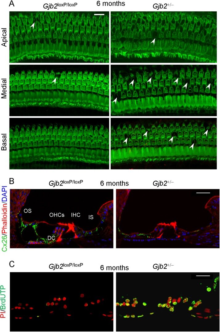
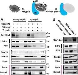
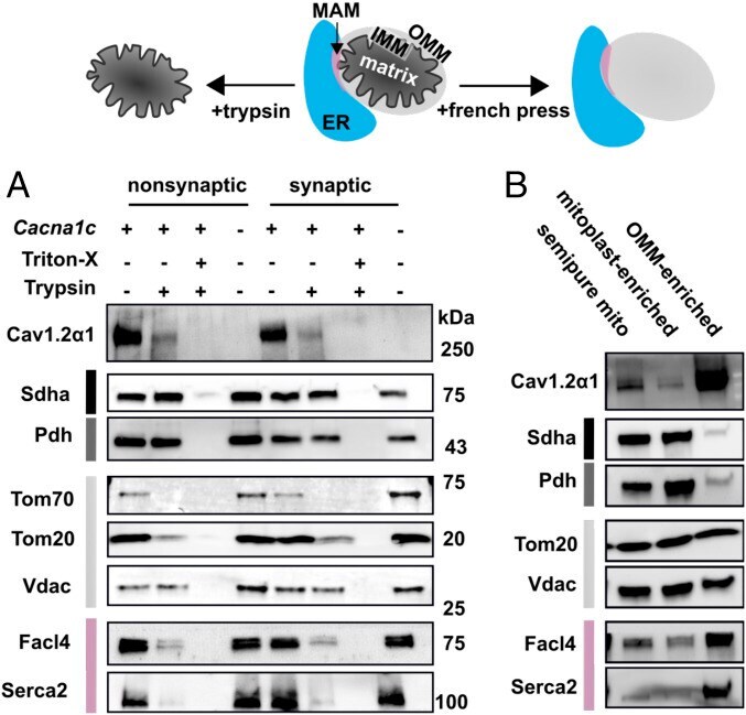
- Fig. 1. Ca v 1.2alpha1 fractionates with OMMs and MAMs in semipure mitochondria. ( A ) Representative immunoblots of submitochondrial fractions before and after trypsin digest in semipure mitochondria isolated from WT (+) and brain-Cacna1c brain-KO (-) mice. Subcellular compartments are color coded as ER (blue), MAM-enriched (pink), OMM (light gray), IMM (black), and mitochondrial matrix (dark gray). Trypsin was used to digest synaptic and nonsynaptic mitochondrially enriched fractions; 1% Triton-X was used as a control to solubilize all proteins, rendering them accessible to trypsin digest ( n = 4, combined from three experiments). ( B ) Representative immunoblots from French press disrupted mitochondria on nondisrupted mitochondria (semipure mito), mitoplast-enriched fractions, and OMM-enriched cell fractions ( n = 8, combined from three experiments).
- Submitted by
- Invitrogen Antibodies (provider)
- Main image
- Experimental details
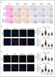
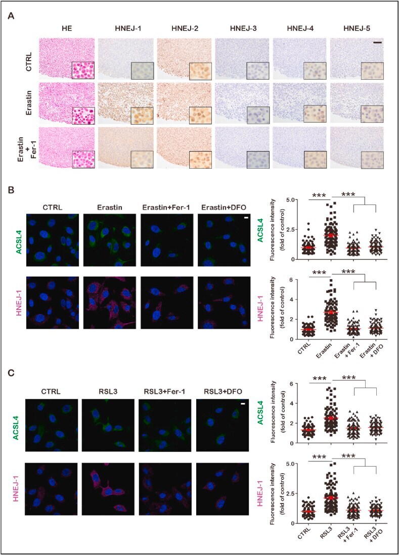
- Fig. 1 Identification of HNEJ-1 as a ferroptosis-specific antibody among five monoclonal anti-HNE antibodies. (A) Human fibrosarcoma HT1080 cells were treated with erastin (10 muM) in the presence or absence of ferrostatin-1 (Fer-1, 5 muM) for 12 h. Then, they were harvested and fixed with 4% paraformaldehyde (PFA) for cell block. Immunohistochemistry was performed with HNEJ-1~5, respectively (scale bar = 100 mum). (B) Human fibrosarcoma HT1080 cells were treated with erastin (10 muM) in the presence or absence of Fer-1 (5 muM) or deferoxamine mesylate (DFO, 500 muM) for 12 h. Then, they were fixed with 4% PFA. After blocking, cells were incubated with HNEJ-1 or anti-ACSL4 and finally observed with confocal microscopy (scale bar = 10 mum). (C) Human fibrosarcoma HT1080 cells were treated with RSL3 (0.25 muM) in the presence or absence of Fer-1 (5 muM) or DFO (500 muM) for 3 h. Then, they were fixed with 4% PFA. After blocking, cells were then incubated with HNEJ-1 or anti-ACSL4 antibody and finally observed with confocal microscopy (scale bar = 10 mum). Representative data are shown based on 3 independent experiments and the analysis is shown as means +- SEM (n = 3); ***P < 0.001 vs control (CTRL) unless indicated by bar. Refer to text for details. Fig. 1
- Submitted by
- Invitrogen Antibodies (provider)
- Main image
- Experimental details
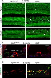
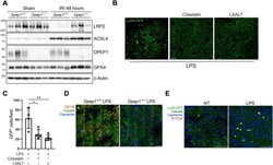
- Fig. 7. DPEP1-mediated leukocyte recruitment and tubular injury. ( A ) Immunoblotting for LRP2, GPX4, ACSL4, and DPEP1 expression in whole-kidney tissue from Dpep1 -/- and Dpep1 +/+ mice 48 hours after renal IRI or sham operation. ( B ) Kidney IVM in LysM gfp/gfp mice at 90 min following LPS administration with or without cilastatin or LSALT peptide treatment. Scale bars, 100 mum. ( C ) Stationary GFP + leukocytes/field in the kidney were quantified (versus LPS: cilastatin, * P = 0.01; LSALT, ** P = 0.002; n = 4 to 5 per group, ANOVA with Dunnett's post hoc test). ( D ) Kidney IVM in Dpep1 +/+ and Dpep1 -/- mice at 90 min following LPS administration. Labels: leukocytes (CD11b, red), capillaries (QTracker, blue), and tubules (autofluorescence, yellow-green). Scale bars, 100 mum. ( E ) Kidney IVM with SYTOX Red staining in LysM gfp/gfp mice 2 hours following LPS administration. Non-LPS (NT)-treated mice are shown as a control. Labels: leukocytes (LysM-GFP, bright green/yellow), tubules (autofluorescence, dark green), capillaries (QTracker, blue), and necrotic cells (SYTOX, red). Scale bars, 100 mum.
