 Explore
 Validate
 Learn
Learn Western blot
 Immunocytochemistry
Antibody data
- Antibody Data
- Antigen structure
- References [1]
- Comments [0]
- Validations
- Immunocytochemistry [1]
- Immunohistochemistry [1]
Submit
Validation data
Reference
Comment
Report error
- Product number
- HPA019824 - Provider product page

- Provider
- Atlas Antibodies
- Proper citation
- Atlas Antibodies Cat#HPA019824, RRID:AB_1856113
- Product name
- Anti-RBM15
- Antibody type
- Polyclonal
- Description
- Polyclonal Antibody against Human RBM15, Gene description: RNA binding motif protein 15, Alternative Gene Names: OTT, OTT1, Validated applications: WB, IHC, ICC, Uniprot ID: Q96T37, Storage: Store at +4°C for short term storage. Long time storage is recommended at -20°C.
- Reactivity
- Human
- Host
- Rabbit
- Conjugate
- Unconjugated
- Isotype
- IgG
- Vial size
- 100 µl
- Concentration
- 0.1 mg/ml
- Storage
- Store at +4°C for short term storage. Long time storage is recommended at -20°C.
- Handling
- The antibody solution should be gently mixed before use.
Submitted references METTL3/MYCN cooperation drives neural crest differentiation and provides therapeutic vulnerability in neuroblastoma.
Thombare K, Vaid R, Pucci P, Ihrmark Lundberg K, Ayyalusamy R, Baig MH, Mendez A, Burgos-Panadero R, Höppner S, Bartenhagen C, Sjövall D, Rehan AA, Dattatraya Nale S, Djos A, Martinsson T, Jaako P, Dong JJ, Kogner P, Johnsen JI, Fischer M, Turner SD, Mondal T
The EMBO journal 2024 Dec;43(24):6310-6335
The EMBO journal 2024 Dec;43(24):6310-6335
No comments: Submit comment
Supportive validation
- Submitted by
- Atlas Antibodies (provider)
- Main image
- Experimental details
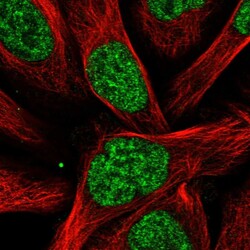
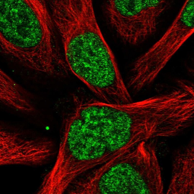
- Immunofluorescent staining of human cell line U-2 OS shows localization to nucleoplasm.
- Sample type
- Human
Supportive validation
- Submitted by
- Atlas Antibodies (provider)
- Enhanced method
- Orthogonal validation
- Main image
- Experimental details
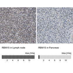
- Immunohistochemistry analysis in human lymph node and pancreas tissues using HPA019824 antibody. Corresponding RBM15 RNA-seq data are presented for the same tissues.
- Sample type
- Human
- Protocol
- Protocol
