Explore
 Validate
 Learn
Learn Western blot
 Immunohistochemistry
Antibody data
- Antibody Data
- Antigen structure
- References [4]
- Comments [0]
- Validations
- Immunohistochemistry [4]
Submit
Validation data
Reference
Comment
Report error
- Product number
- NBP1-87748 - Provider product page

- Provider
- Novus Biologicals
- Proper citation
- Novus Cat#NBP1-87748, RRID:AB_11035627
- Product name
- Rabbit Polyclonal myosin X Antibody
- Antibody type
- Polyclonal
- Description
- Immunogen affinity purified. Specificity of human myosin X antibody verified on a Protein Array containing target protein plus 383 other non-specific proteins.
- Reactivity
- Human
- Host
- Rabbit
- Isotype
- IgG
- Vial size
- 0.1 ml
- Storage
- Store at 4C short term. Aliquot and store at -20C long term. Avoid freeze-thaw cycles.
Submitted references DPP6 regulation of dendritic morphogenesis impacts hippocampal synaptic development.
Myosin X and its motorless isoform differentially modulate dendritic spine development by regulating trafficking and retention of vasodilator-stimulated phosphoprotein.
Myosin-X functions in polarized epithelial cells.
Headless Myo10 is a negative regulator of full-length Myo10 and inhibits axon outgrowth in cortical neurons.
Lin L, Sun W, Throesch B, Kung F, Decoster JT, Berner CJ, Cheney RE, Rudy B, Hoffman DA
Nature communications 2013;4:2270
Nature communications 2013;4:2270
Myosin X and its motorless isoform differentially modulate dendritic spine development by regulating trafficking and retention of vasodilator-stimulated phosphoprotein.
Lin WH, Hurley JT, Raines AN, Cheney RE, Webb DJ
Journal of cell science 2013 Oct 15;126(Pt 20):4756-68
Journal of cell science 2013 Oct 15;126(Pt 20):4756-68
Myosin-X functions in polarized epithelial cells.
Liu KC, Jacobs DT, Dunn BD, Fanning AS, Cheney RE
Molecular biology of the cell 2012 May;23(9):1675-87
Molecular biology of the cell 2012 May;23(9):1675-87
Headless Myo10 is a negative regulator of full-length Myo10 and inhibits axon outgrowth in cortical neurons.
Raines AN, Nagdas S, Kerber ML, Cheney RE
The Journal of biological chemistry 2012 Jul 20;287(30):24873-83
The Journal of biological chemistry 2012 Jul 20;287(30):24873-83
No comments: Submit comment
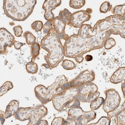
Supportive validation
- Submitted by
- Novus Biologicals (provider)
- Main image
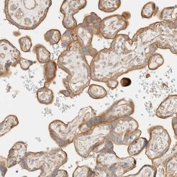
- Experimental details
- Immunohistochemistry-Paraffin: myosin X Antibody [NBP1-87748] - Staining of human placenta shows moderate cytoplasmic positivity in trophoblastic cells.
- Submitted by
- Novus Biologicals (provider)
- Main image
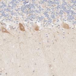
- Experimental details
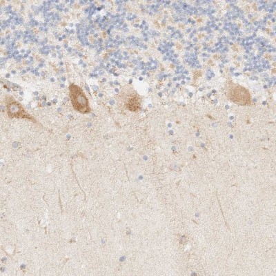
- Immunohistochemistry-Paraffin: myosin X Antibody [NBP1-87748] - Staining of human cerebellum shows moderate cytoplasmic positivity in Purkinje cells.
- Submitted by
- Novus Biologicals (provider)
- Main image
- Experimental details
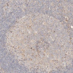
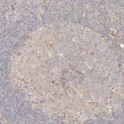
- Immunohistochemistry-Paraffin: myosin X Antibody [NBP1-87748] - Staining of human lymphoid tissues shows weak cytoplasmic positivity in germinal center cells.
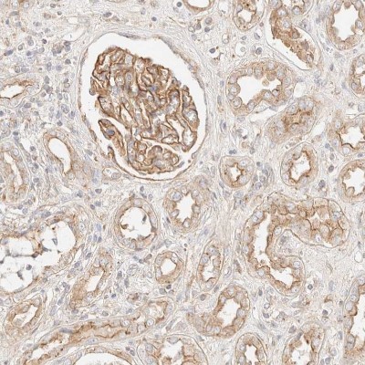
- Submitted by
- Novus Biologicals (provider)
- Main image

- Experimental details
- Immunohistochemistry-Paraffin: myosin X Antibody [NBP1-87748] - Staining of human kidney shows strong membranous positivity in cells in glomeruli.