Explore
 Validate
 Learn
Learn Western blot
Antibody data
- Antibody Data
- Antigen structure
- References [0]
- Comments [0]
- Validations
- Western blot [2]
- ELISA [1]
- Immunocytochemistry [1]
- Immunohistochemistry [5]
Submit
Validation data
Reference
Comment
Report error
- Product number
- TA590555 - Provider product page

- Provider
- OriGene
- Product name
- Rabbit Polyclonal PROSC Antibody
- Antibody type
- Polyclonal
- Description
- Rabbit Polyclonal PROSC Antibody
- Host
- Rabbit
- Conjugate
- Unconjugated
- Epitope
- PLPBP
- Isotype
- IgG
- Antibody clone number
- NULL
- Vial size
- 100 µg
- Concentration
- 1.25mg/ml
No comments: Submit comment
Supportive validation
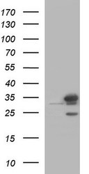
- Submitted by
- OriGene (provider)
- Main image
- Experimental details
- Western Blot: PROSC Antibody - Samples: Lane 1, Marker: 250, 130, 95, 72, 55, 36, 28, 17, 11 Lane 2, RT-4 Lane 3, U-251MG sp Lane 4, Human Plasma Lane 5, Liver Lane 6, Tonsil , Target weight: 30 Validation score: 2 Validation description: Supportive - Band of predicted size in kDa (+/-20%) with additional bands present.
- Validation comment
- WB

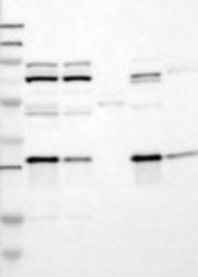
- Submitted by
- OriGene (provider)
- Main image
- Experimental details
- HEK293T cells were transfected with the pCMV6-ENTRY control (Left lane) or pCMV6-ENTRY PROSC (RC200853, Right lane) cDNA for 48 hrs and lysed. Equivalent amounts of cell lysates (5 ug per lane) were separated by SDS-PAGE and immunoblotted with anti-PROSC.
- Validation comment
- WB

Supportive validation
- Submitted by
- OriGene (provider)
- Main image
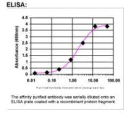
- Experimental details
- ELISA: PROSC Antibody
- Validation comment
- ELISA

Supportive validation
- Submitted by
- OriGene (provider)
- Main image
- Experimental details
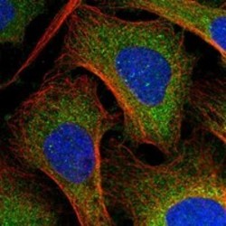
- Immunofluorescent staining of human cell line U-2 OS shows positivity in cytoplasm. Antibody dilution: 1:75. Image and statement courtesy of the Human Protein Atlas (HPA).
- Validation comment
- IF

Supportive validation
- Submitted by
- OriGene (provider)
- Main image
- Experimental details
- Immunohistochemical staining of human small intestine shows strong cytoplasmic positivity in glandular cells. Antibody dilution: 1:500. Image and statement courtesy of the Human Protein Atlas (HPA).
- Validation comment
- IHC
- Submitted by
- OriGene (provider)
- Main image
- Experimental details
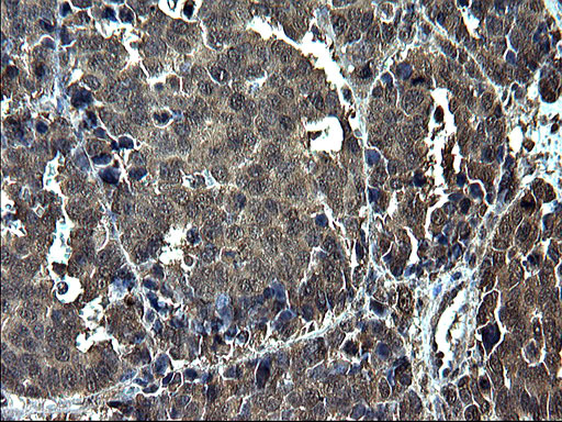
- Immunohistochemical staining of paraffin-embedded Adenocarcinoma of Human colon tissue using anti-PROSC Rabbit Polyclonal antibody. (TA590555)
- Validation comment
- IHC
- Submitted by
- OriGene (provider)
- Main image
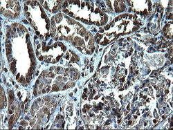
- Experimental details
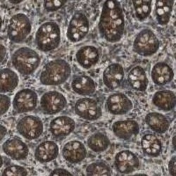
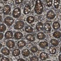
- Immunohistochemical staining of paraffin-embedded Human Kidney tissue using anti-PROSC Rabbit Polyclonal antibody. (TA590555)
- Validation comment
- IHC
- Submitted by
- OriGene (provider)
- Main image

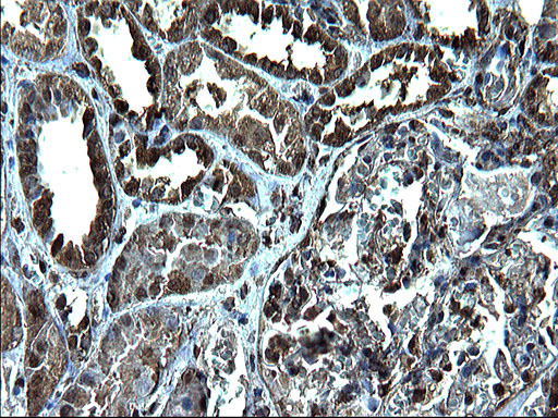
- Experimental details
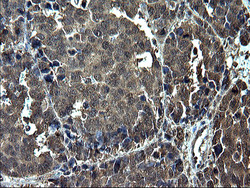
- Immunohistochemical staining of paraffin-embedded Carcinoma of Human liver tissue using anti-PROSC Rabbit Polyclonal antibody. (TA590555)
- Validation comment
- IHC
- Submitted by
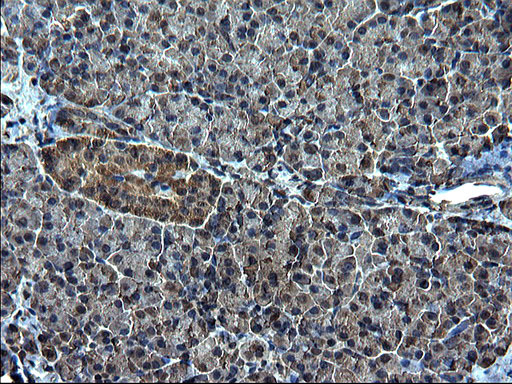
- OriGene (provider)
- Main image
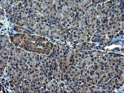
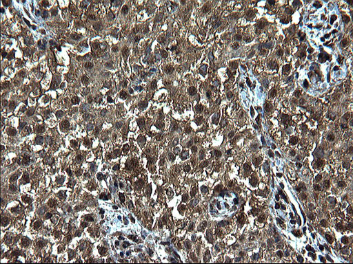
- Experimental details
- Immunohistochemical staining of paraffin-embedded Human pancreas tissue using anti-PROSC Rabbit Polyclonal antibody. (TA590555)
- Validation comment
- IHC