Explore
 Validate
 Learn
Learn Western blot
 Immunohistochemistry
Antibody data
- Antibody Data
- Antigen structure
- References [3]
- Comments [0]
- Validations
- Immunohistochemistry [1]
Submit
Validation data
Reference
Comment
Report error
- Product number
- GTX22868 - Provider product page

- Provider
- GeneTex
- Proper citation
- GeneTex Cat#GTX22868, RRID:AB_384887
- Product name
- Ryanodine Receptor antibody [34C]
- Antibody type
- Monoclonal
- Reactivity
- Human, Mouse, Rat, Bovine, Canine, Chicken/Avian, Porcine, Rabbit, Sheep, Simian
- Host
- Mouse
Submitted references Three-dimensional reconstruction of the recombinant type 3 ryanodine receptor and localization of its amino terminus.
Presenilin-1 mutations increase levels of ryanodine receptors and calcium release in PC12 cells and cortical neurons.
Long-chain acylcarnitine induces Ca2+ efflux from the sarcoplasmic reticulum.
Liu Z, Zhang J, Sharma MR, Li P, Chen SR, Wagenknecht T
Proceedings of the National Academy of Sciences of the United States of America 2001 May 22;98(11):6104-9
Proceedings of the National Academy of Sciences of the United States of America 2001 May 22;98(11):6104-9
Presenilin-1 mutations increase levels of ryanodine receptors and calcium release in PC12 cells and cortical neurons.
Chan SL, Mayne M, Holden CP, Geiger JD, Mattson MP
The Journal of biological chemistry 2000 Jun 16;275(24):18195-200
The Journal of biological chemistry 2000 Jun 16;275(24):18195-200
Long-chain acylcarnitine induces Ca2+ efflux from the sarcoplasmic reticulum.
Yamada KA, Kanter EM, Newatia A
Journal of cardiovascular pharmacology 2000 Jul;36(1):14-21
Journal of cardiovascular pharmacology 2000 Jul;36(1):14-21
No comments: Submit comment
Supportive validation
- Submitted by
- GeneTex (provider)
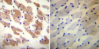
- Main image

- Experimental details
- Immunohistochemistry was performed on biopsies of deparaffinized Human skeletal muscle tissue. To expose target proteins, heat induced antigen retrieval was performed using 10mM sodium citrate (pH 6.0) buffer, microwaved for 8-15 minutes. Following antigen retrieval tissues were blocked in 3% BSA-PBS for 30 minutes at room temperature. Tissues were then probed at a dilution of 1:20 with a mouse monoclonal antibody recognizing Ryanodine Receptor (GTX22868) or without primary antibody (negative control) overnight at 4°C in a humidified chamber. Tissues were washed extensively with PBST and endogenous peroxidase activity was quenched with a peroxidase suppressor. Detection was performed using a biotin-conjμgated secondary antibody and SA-HRP, followed by colorimetric detection using DAB. Tissues were counterstained with hematoxylin and prepped for mounting.