 Explore
 Validate
 Learn
Learn Western blot
Antibody data
- Antibody Data
- Antigen structure
- References [0]
- Comments [0]
- Validations
- Western blot [1]
- ELISA [1]
- Immunocytochemistry [1]
- Immunohistochemistry [4]
Submit
Validation data
Reference
Comment
Report error
- Product number
- TA590763 - Provider product page

- Provider
- OriGene
- Product name
- Rabbit Polyclonal LSP1 Antibody
- Antibody type
- Polyclonal
- Description
- Rabbit Polyclonal LSP1 Antibody
- Host
- Rabbit
- Conjugate
- Unconjugated
- Epitope
- LSP1
- Isotype
- IgG
- Antibody clone number
- NULL
- Vial size
- 100 µg
- Concentration
- 0.84mg/ml
No comments: Submit comment
Supportive validation
- Submitted by
- OriGene (provider)
- Main image

- Experimental details
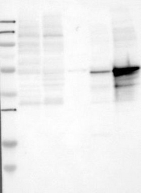
- Lane 1: Marker [kDa] 250, 130, 95, 72, 55, 36, 28, 17, 11; Lane 2: RT-4; Lane 3: U-251 MG; Lane 4: Human Plasma; Lane 5: Liver; Lane 6: TonsilThis validation was performed by Protein Atlas and the presentation of data is for informational purposes only.
- Validation comment
- WB
Supportive validation
- Submitted by
- OriGene (provider)
- Main image
- Experimental details
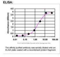
- ELISA: LSP1 Antibody
- Validation comment
- ELISA

Supportive validation
- Submitted by
- OriGene (provider)
- Main image
- Experimental details
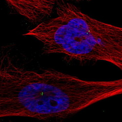
- Immunofluorescent staining of human cell line U-251 MG shows positivity in centrosome.This validation was performed by Protein Atlas and the presentation of data is for informational purposes only.
- Validation comment
- IF

Supportive validation
- Submitted by
- OriGene (provider)
- Main image
- Experimental details
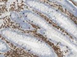
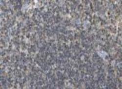
- Immunohistochemistry: LSP1 Antibody - Immunohistochemical staining for lymphocyte-specific protein 1 (LSP1) in human lymphoidal tissues. Lymphoid tissues of appendix displayed moderate to strong cytoplasmic positivity. Normal epithelial and stromal cells were negative. Most malignant lymphomas and a few cases of urothelial cancers expressed moderate cytoplasmic immunoreactivity (images not shown). These image are the courtesy of the Human Proteome Resource (HPR).
- Validation comment
- IHC

- Submitted by
- OriGene (provider)
- Main image
- Experimental details
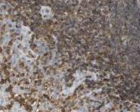
- Immunohistochemistry: LSP1 Antibody - Immunohistochemical staining for lymphocyte-specific protein 1 (LSP1) in human lymphoidal tissues. Lymphoid tissues of lymph nodes displayed moderate to strong cytoplasmic positivity. Normal epithelial and stromal cells were negative. Most malignant lymphomas and a few cases of urothelial cancers expressed moderate cytoplasmic immunoreactivity (images not shown). These image are the courtesy of the Human Proteome Resource (HPR).
- Validation comment
- IHC

- Submitted by
- OriGene (provider)
- Main image

- Experimental details
- Immunohistochemistry: LSP1 Antibody - Immunohistochemical staining for lymphocyte-specific protein 1 (LSP1) in human lymphoidal tissues. Lymphoid tissues of tonsil displayed moderate to strong cytoplasmic positivity. Normal epithelial and stromal cells were negative. Most malignant lymphomas and a few cases of urothelial cancers expressed moderate cytoplasmic immunoreactivity (images not shown). These image are the courtesy of the Human Proteome Resource (HPR).
- Validation comment
- IHC
- Submitted by
- OriGene (provider)
- Main image

- Experimental details
- Immunohistochemical staining of human spleen shows strong cytoplasmic positivity in lymphocytes.This validation was performed by Protein Atlas and the presentation of data is for informational purposes only.
- Validation comment
- IHC
