 Explore
 Validate
 Learn
Learn Western blot
Antibody data
- Antibody Data
- Antigen structure
- References [0]
- Comments [0]
- Validations
- Western blot [1]
- ELISA [1]
- Immunohistochemistry [4]
Submit
Validation data
Reference
Comment
Report error
- Product number
- 24780002-0.1mg - Provider product page

- Provider
- Novus Biologicals
- Product name
- Rabbit Polyclonal LSP1 Antibody
- Antibody type
- Polyclonal
- Description
- Immunogen affinity purified. This product is specific for Human LSP1.
- Reactivity
- Human
- Host
- Rabbit
- Isotype
- IgG
- Vial size
- 0.1 mg
- Storage
- Store at 4C short term. Aliquot and store at -20C long term. Avoid freeze-thaw cycles.
No comments: Submit comment
Supportive validation
- Submitted by
- Novus Biologicals (provider)
- Main image

- Experimental details
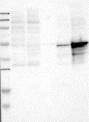
- Western Blot: LSP1 Antibody [24780002] - Samples: Lane 1, Marker [kDa]: 250, 130, 95, 72, 55, 36, 28, 17, 11 Lane 2, RT-4 Lane 3, U-251MG sp Lane 4, Human Plasma Lane 5, Liver Lane 6, Tonsil, Target weight [kDa]: 50, 38, 37, 30 (splice variants) Validation score: 2 Validation description: Supportive - Band of predicted size in kDa (+/-20%) with additional bands present.
Supportive validation
- Submitted by
- Novus Biologicals (provider)
- Main image
- Experimental details
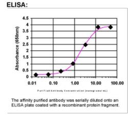
- ELISA: LSP1 Antibody [24780002]

Supportive validation
- Submitted by
- Novus Biologicals (provider)
- Main image

- Experimental details
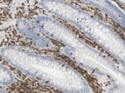
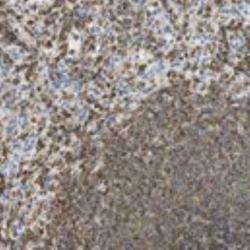
- Immunohistochemistry: LSP1 Antibody [24780002] - Staining for lymphocyte-specific protein 1 (LSP1) in human lymphoidal tissues. Lymphoid tissues of lymph nodes displayed moderate to strong cytoplasmic positivity. Normal epithelial and stromal cells were negative. Most malignant lymphomas and a few cases of urothelial cancers expressed moderate cytoplasmic immunoreactivity (images not shown). These image are the courtesy of the Human Proteome Resource (HPR).

- Submitted by
- Novus Biologicals (provider)
- Main image
- Experimental details
- Immunohistochemistry: LSP1 Antibody [24780002] - staining for lymphocyte-specific protein 1 (LSP1) in human lymphoidal tissues. Lymphoid tissues of appendix displayed moderate to strong cytoplasmic positivity. Normal epithelial and stromal cells were negative. Most malignant lymphomas and a few cases of urothelial cancers expressed moderate cytoplasmic immunoreactivity (images not shown). These image are the courtesy of the Human Proteome Resource (HPR).

- Submitted by
- Novus Biologicals (provider)
- Main image
- Experimental details
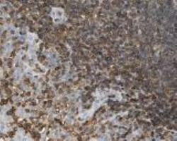
- Immunohistochemistry: LSP1 Antibody [24780002] - Staining for lymphocyte-specific protein 1 (LSP1) in human lymphoidal tissues. Lymphoid tissues of tonsil displayed moderate to strong cytoplasmic positivity. Normal epithelial and stromal cells were negative. Most malignant lymphomas and a few cases of urothelial cancers expressed moderate cytoplasmic immunoreactivity (images not shown). These image are the courtesy of the Human Proteome Resource (HPR).

- Submitted by
- Novus Biologicals (provider)
- Main image
- Experimental details
- Immunohistochemistry: LSP1 Antibody [24780002] - Lymphoid tissues displayed moderate to strong cytoplasmic positivity. Normal epithelial and stromal cells were negative. Among malignant cells, most malignant lymphomas and a few cases of urothelial cancers expressed moderate cytoplasmic immunoreactivity. Image and statement courtesy of the Human Protein Atlas (HPA).
