Explore
 Validate
 Learn
Learn Western blot
 Immunoprecipitation
 Chromatin Immunoprecipitation
Antibody data
- Antibody Data
- Antigen structure
- References [1]
- Comments [0]
- Validations
- Immunoprecipitation [1]
- Immunohistochemistry [2]
- Other assay [3]
Submit
Validation data
Reference
Comment
Report error
- Product number
- PA5-30586 - Provider product page

- Provider
- Invitrogen Antibodies
- Product name
- TAL1 Polyclonal Antibody
- Antibody type
- Polyclonal
- Antigen
- Synthetic peptide
- Description
- Recommended positive controls: H1299. Predicted reactivity: Bovine (100%). Store product as a concentrated solution. Centrifuge briefly prior to opening the vial.
- Reactivity
- Human, Mouse
- Host
- Rabbit
- Isotype
- IgG
- Vial size
- 100 μL
- Concentration
- 1.61 mg/mL
- Storage
- Store at 4°C short term. For long term storage, store at -20°C, avoiding freeze/thaw cycles.
Submitted references Dengue virus infection impedes megakaryopoiesis in MEG-01 cells where the virus envelope protein interacts with the transcription factor TAL-1.
Banerjee A, Tripathi A, Duggal S, Banerjee A, Vrati S
Scientific reports 2020 Nov 11;10(1):19587
Scientific reports 2020 Nov 11;10(1):19587
No comments: Submit comment
Supportive validation
- Submitted by
- Invitrogen Antibodies (provider)
- Main image
- Experimental details
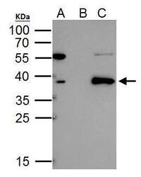
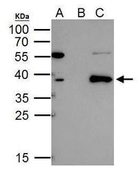
- TAL1 antibody immunoprecipitates TAL1 protein in IP experiments. IP Sample: 293T whole cell lysate/extract A. 40 µg 293T whole cell lysate/extract B. Control with 2 µg of preimmune rabbit IgG C. Immunoprecipitation of TAL1 protein by 2 µg of TAL1 antibody (Product # PA5-30586) 12% SDS-PAGE The immunoprecipitated TAL1 protein was detected by TAL1 antibody (Product # PA5-30586) diluted at 1:1,000.
Supportive validation
- Submitted by
- Invitrogen Antibodies (provider)
- Main image
- Experimental details
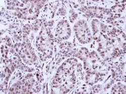
- TAL1 Polyclonal Antibody detects TAL1 protein at cytosol and nucleus on mouse spleen by immunohistochemical analysis. Sample: Paraffin-embedded mouse spleen. TAL1 Polyclonal Antibody (Product # PA5-30586) dilution: 1:500. Antigen Retrieval: EDTA based buffer, pH 8.0, 15 min.

- Submitted by
- Invitrogen Antibodies (provider)
- Main image
- Experimental details
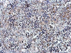
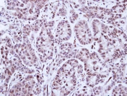
- Immunohistochemical analysis of paraffin-embedded A549 Xenograft , using TAL1 (Product # PA5-30586) antibody at 1:100 dilution. Antigen Retrieval: EDTA based buffer, pH 8.0, 15 min.
Supportive validation
- Submitted by
- Invitrogen Antibodies (provider)
- Main image
- Experimental details
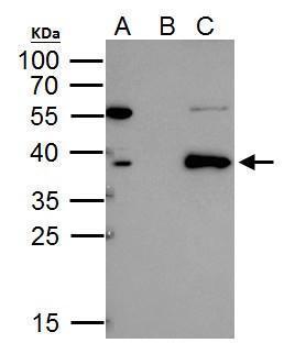
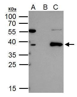
- TAL1 antibody immunoprecipitates TAL1 protein in IP experiments. IP Sample: 293T whole cell lysate/extract A. 40 µg 293T whole cell lysate/extract B. Control with 2 µg of preimmune rabbit IgG C. Immunoprecipitation of TAL1 protein by 2 µg of TAL1 antibody (Product # PA5-30586) 12% SDS-PAGE The immunoprecipitated TAL1 protein was detected by TAL1 antibody (Product # PA5-30586) diluted at 1:1,000.
- Submitted by
- Invitrogen Antibodies (provider)
- Main image
- Experimental details
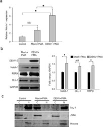
- Figure 5 Notch signaling in DENV-infected MEG-01 cells. Naive MEG-01 cells (control) were treated with PMA (Mock + PMA), or DENV-infected (MOI-1) cells were treated with PMA (DENV + PMA). Cells were harvested at day 5 pi for isolating total RNA and preparation of cell lysate. ( a ) Expression of Notch-1 transcript relative to Gapdh expression is presented from 3 independent experiments. ( b ) The cell lysate was Western blotted for different proteins (left panel) and the band intensities were quantified by ImageJ software. Intensities from 3 or more blots were determined compared to GAPDH, and average intensities in DENV + PMA compared to Mock + PMA treatment were plotted to show the fold-change in protein expression (right panel). Intensities from 3 or more blots were determined. ( c ) Nuclear (N) and cytoplasmic (C) extracts of MEG-01 cells were prepared and Western blotted with TAL-1 antibody. Levels of Actin and Histone proteins were checked to demonstrate the quality of protein fractionation. Statistical analyses were done using the one-way analysis of variance (ANOVA). * p < 0.05, ** p < 0.005, NS denotes difference not-significant.

- Submitted by
- Invitrogen Antibodies (provider)
- Main image

- Experimental details
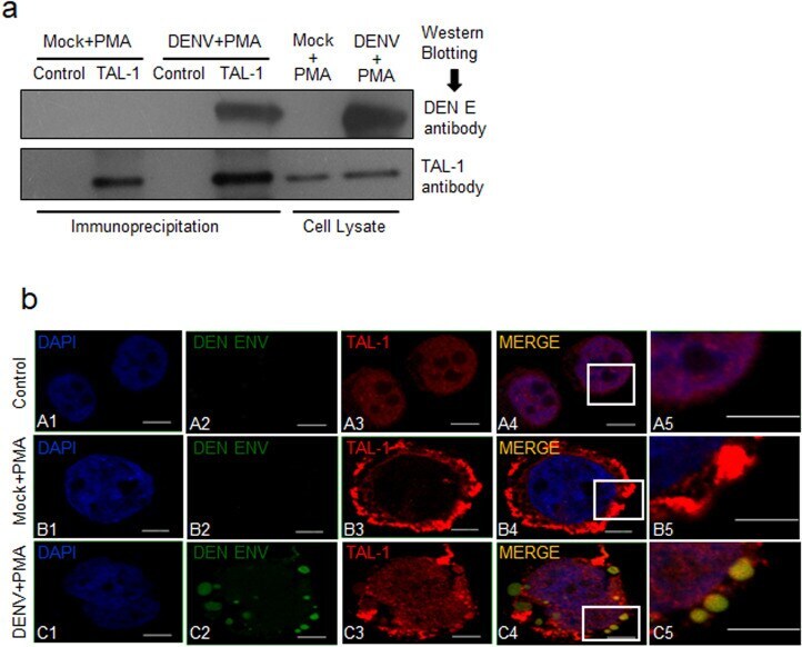
- Figure 6 Interaction of Tal-1 and DENV E protein. Naive MEG-01 cells (control) were treated with PMA (Mock + PMA), or DENV-infected (MOI-1) cells were treated with PMA (DENV + PMA). ( a ) Cells were harvested at day 5 pi for preparation of cell lysate. Immunoprecipitation was carried out using the rabbit anti-TAL-1 antibody, or the rabbit IgG as the isotype control, and the Protein A/G magnetic beads. The precipitated proteins and the cell lysate used for the immunoprecipitation were Western blotted with antibodies shown on the right side of the figure. ( b ) On day 5 pi, cells were harvested, fixed in 2% paraformaldehyde, followed by permeabilization with 0.03% Triton-X. This was followed by incubation with TAL-1, and DENV E antibodies and stained with the fluorescent-labeled secondary antibody. Cells were mounted on slides using ProLong Gold anti-fade reagent with DAPI and images taken using a confocal microscope. Untreated MEG-01 cells were used as a control. Experiments were performed 3 times and representative confocal images are shown. The right-most panels are zoomed up images of the insets in the adjacent panels. The scale bar size is 5 mum.