Explore
 Validate
 Learn
Learn Immunohistochemistry
Antibody data
- Antibody Data
- Antigen structure
- References [0]
- Comments [0]
- Validations
- Immunohistochemistry [3]
Submit
Validation data
Reference
Comment
Report error
- Product number
- PA5-46968 - Provider product page

- Provider
- Invitrogen Antibodies
- Product name
- Angiopoietin 1 Polyclonal Antibody
- Antibody type
- Polyclonal
- Antigen
- Recombinant full-length protein
- Description
- In direct ELISAs, less than 2% cross-reactivity with recombinant human (rh) Angiopoietin-2, rhAngiopoietin-4, and rhAngiopoietin-like Factor/CDT6 is observed.
- Concentration
- 0.2 mg/mL
No comments: Submit comment
Supportive validation
- Submitted by
- Invitrogen Antibodies (provider)
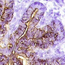
- Main image

- Experimental details
- Immunohistochemical analysis of Angiopoietin 1 in immersion fixed paraffin-embedded sections of human prostate cancer tissue. Samples were incubated in Angiopoietin 1 polyclonal antibody (Product # PA5-46968) using a dilution of 15 µg/mL overnight at 4 °C. Tissue was stained with the Anti-Goat HRP-DAB Cell & Tissue Staining Kit (brown) and counterstained with hematoxylin (blue).
- Submitted by
- Invitrogen Antibodies (provider)
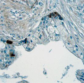
- Main image
- Experimental details
- Immunohistochemical analysis of Angiopoietin 1 in perfusion fixed frozen sections of mouse embryo (15 d.p.c.) . Samples were incubated in Angiopoietin 1 polyclonal antibody (Product # PA5-46968) using a dilution of 15 µg/mL overnight at 4 °C. Tissue was stained using the Anti-Goat HRP-DAB Cell & Tissue Staining Kit (brown) and counterstained with hematoxylin (blue). Specific staining was localized to endothelium in the loop of the midgut.

- Submitted by
- Invitrogen Antibodies (provider)
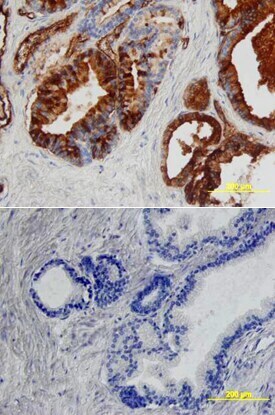
- Main image

- Experimental details
- Immunohistochemical analysis of Angiopoietin 1 in immersion fixed paraffin-embedded sections of human prostate array. Samples were incubated in Angiopoietin 1 polyclonal antibody (Product # PA5-46968) using a dilution of 5 µg/mL overnight at 4 °C. Tissue was stained using the Anti-Goat HRP-DAB Cell & Tissue Staining Kit (brown) and counterstained with hematoxylin (blue). Lower panel shows a lack of labeling if primary antibodies are omitted and tissue is stained only with secondary antibody followed by incubation with detection reagents.