Explore
 Validate
 Learn
Learn Immunoprecipitation
Antibody data
- Antibody Data
- Antigen structure
- References [3]
- Comments [0]
- Validations
- Immunoprecipitation [1]
- Immunohistochemistry [1]
Submit
Validation data
Reference
Comment
Report error
- Product number
- PAB10092 - Provider product page

- Provider
- Abnova Corporation
- Proper citation
- Abnova Corporation Cat#PAB10092, RRID:AB_1671894
- Product name
- Angpt1 polyclonal antibody
- Antibody type
- Polyclonal
- Description
- Rabbit polyclonal antibody raised against synthetic peptide of Angpt1.
- Storage
- Store at 4°C. For long term storage store at -20°C.Aliquot to avoid repeated freezing and thawing.
Submitted references Angiopoietin-2, a natural antagonist for Tie2 that disrupts in vivo angiogenesis.
Requisite role of angiopoietin-1, a ligand for the TIE2 receptor, during embryonic angiogenesis.
Isolation of angiopoietin-1, a ligand for the TIE2 receptor, by secretion-trap expression cloning.
Maisonpierre PC, Suri C, Jones PF, Bartunkova S, Wiegand SJ, Radziejewski C, Compton D, McClain J, Aldrich TH, Papadopoulos N, Daly TJ, Davis S, Sato TN, Yancopoulos GD
Science (New York, N.Y.) 1997 Jul 4;277(5322):55-60
Science (New York, N.Y.) 1997 Jul 4;277(5322):55-60
Requisite role of angiopoietin-1, a ligand for the TIE2 receptor, during embryonic angiogenesis.
Suri C, Jones PF, Patan S, Bartunkova S, Maisonpierre PC, Davis S, Sato TN, Yancopoulos GD
Cell 1996 Dec 27;87(7):1171-80
Cell 1996 Dec 27;87(7):1171-80
Isolation of angiopoietin-1, a ligand for the TIE2 receptor, by secretion-trap expression cloning.
Davis S, Aldrich TH, Jones PF, Acheson A, Compton DL, Jain V, Ryan TE, Bruno J, Radziejewski C, Maisonpierre PC, Yancopoulos GD
Cell 1996 Dec 27;87(7):1161-9
Cell 1996 Dec 27;87(7):1161-9
No comments: Submit comment
Supportive validation
- Submitted by
- Abnova Corporation (provider)
- Main image
- Experimental details
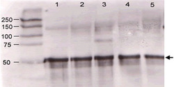
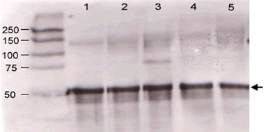
- Immunoblotting of Angpt1 polyclonal antibody (Cat # PAB10092) was used at a 1:500 dilution to detect mouse Angptl1 by western blot against supernatants of mouse angiopoietin-expressing endothelial cells.Lane 1 - wt endothelial cells.Lane 2 - mouse Angpt1 (clone 1-8) expressing cells.Lane 3 - mouse Angpt1 (clone 1-15) expressing cells.Lane 4 - mouse Angpt2 (clone 2-9) expressing cells.Approximately 20 ug of each lysate was used for 10% SDS-PAGE.Immunoprecipitation preceded the reaction with primary antibody at room temperature for 1 h.After subsequent washing, a 1:5,000 dilution of HRP conjugated Gt-a-Rabbit IgG preceded color development.
- Validation comment
- Immunoprecipitation
Supportive validation
- Submitted by
- Abnova Corporation (provider)
- Main image

- Experimental details
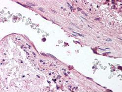
- Immunohistochemical staining with Angpt1 polyclonal antibody (Cat # PAB10092) was diluted 1 : 500 to detect Angptl1 in human lung tissue. Tissue was formalin fixed and paraffin embedded. No pre-treatment of sample was required. The image shows the localization of antibody as the precipitated red signal, with a hematoxylin purple nuclear counter stain.
- Validation comment
- Immunohistochemistry (Formalin/PFA-fixed paraffin-embedded sections)