 Explore
 Validate
 Learn
Learn Western blot
Antibody data
- Antibody Data
- Antigen structure
- References [0]
- Comments [0]
- Validations
- Western blot [2]
- Immunocytochemistry [4]
- Immunohistochemistry [1]
- Flow cytometry [2]
Submit
Validation data
Reference
Comment
Report error
- Product number
- PA5-142604 - Provider product page

- Provider
- Invitrogen Antibodies
- Product name
- Angiopoietin 1 Polyclonal Antibody
- Antibody type
- Polyclonal
- Antigen
- Synthetic peptide
- Description
- This antibody is tested in Peptide ELISA: antibody detection limit dilution 1:32,000.
- Reactivity
- Human
- Host
- Goat
- Isotype
- IgG
- Vial size
- 100 μg
- Concentration
- 0.5 mg/mL
- Storage
- -20°C, Avoid Freeze/Thaw Cycles
No comments: Submit comment
Supportive validation
- Submitted by
- Invitrogen Antibodies (provider)
- Main image
- Experimental details
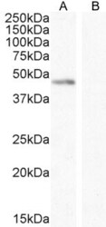
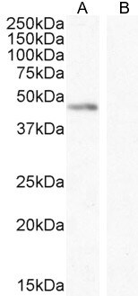
- Western Blot analysis of Angiopoietin 1 in K56 cell lysate (A) + peptide (B) (35 µg protein in RIPA buffer) using Angiopoietin 1 Polyclonal Antibody (Product # PA5-142604) at 0.1 µg/mL. Detected by chemiluminescence.
- Submitted by
- Invitrogen Antibodies (provider)
- Main image
- Experimental details
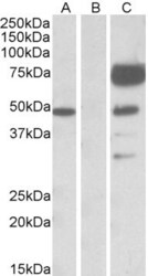
- Western Blot analysis of Angiopoietin 1 in HEK293 lysate (10 µg protein in RIPA buffer) using Angiopoietin 1 Polyclonal Antibody (Product # PA5-142604) at 10 µg protein in RIPA buffer) overexpressing Human ANGPT1. Lane A: HEK293 lysate over expressing Human Angiopoietin 1 with DYKDDDDK tag probed with Angiopoietin 1 Antibody. Lane B: Mock-transfected HEK293 probed with Angiopoietin 1 Antibody. Lane C: HEK293 lysate over expressing Human Angiopoietin 1 with DYKDDDDK tag probed with anti-DYKDDDDK Tag (1:1,000). Primary incubations were for 1 hour. Detected by chemiluminescence.

Supportive validation
- Submitted by
- Invitrogen Antibodies (provider)
- Main image
- Experimental details
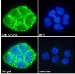
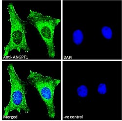
- Immunocytochemistry-Immunofluorescence analysis of Angiopoietin 1 in A431 cells using Angiopoietin 1 Polyclonal Antibody (Product # PA5-142604). Cells were fixed with paraformaldehyde, permeabilized with 0.15% Triton, and incubated with the primary antibody for 1hr (10 µg/mL) followed by Alexa Fluor 488 secondary antibody (2 µg/mL), showing plasma membrane staining. The nuclear stain is DAPI (blue). Negative control: Unimmunized goat IgG (10 µg/mL) followed by Alexa Fluor 488 secondary antibody (2 µg/mL).

- Submitted by
- Invitrogen Antibodies (provider)
- Main image
- Experimental details
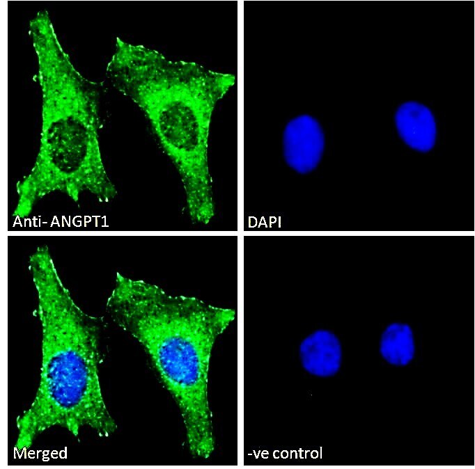
- Immunocytochemistry-Immunofluorescence analysis of Angiopoietin 1 in HeLa cells using Angiopoietin 1 Polyclonal Antibody (Product # PA5-142604). Cells were fixed with paraformaldehyde, permeabilized with 0.15% Triton, and incubated with the primary antibody for 1hr (10 µg/mL) followed by Alexa Fluor 488 secondary antibody (2 µg/mL), showing plasma membrane and cytoplasmic staining. The nuclear stain is DAPI (blue). Negative control: Unimmunized goat IgG (10 µg/mL) followed by Alexa Fluor 488 secondary antibody (2 µg/mL).
- Submitted by
- Invitrogen Antibodies (provider)
- Main image

- Experimental details
- Immunocytochemistry-Immunofluorescence analysis of Angiopoietin 1 in A431 cells using Angiopoietin 1 Polyclonal Antibody (Product # PA5-142604). Cells were fixed with paraformaldehyde, permeabilized with 0.15% Triton, and incubated with the primary antibody for 1hr (10 µg/mL) followed by Alexa Fluor 488 secondary antibody (2 µg/mL), showing plasma membrane staining. The nuclear stain is DAPI (blue). Negative control: Unimmunized goat IgG (10 µg/mL) followed by Alexa Fluor 488 secondary antibody (2 µg/mL).

- Submitted by
- Invitrogen Antibodies (provider)
- Main image

- Experimental details
- Immunocytochemistry-Immunofluorescence analysis of Angiopoietin 1 in HeLa cells using Angiopoietin 1 Polyclonal Antibody (Product # PA5-142604). Cells were fixed with paraformaldehyde, permeabilized with 0.15% Triton, and incubated with the primary antibody for 1hr (10 µg/mL) followed by Alexa Fluor 488 secondary antibody (2 µg/mL), showing plasma membrane and cytoplasmic staining. The nuclear stain is DAPI (blue). Negative control: Unimmunized goat IgG (10 µg/mL) followed by Alexa Fluor 488 secondary antibody (2 µg/mL).

Supportive validation
- Submitted by
- Invitrogen Antibodies (provider)
- Main image

- Experimental details
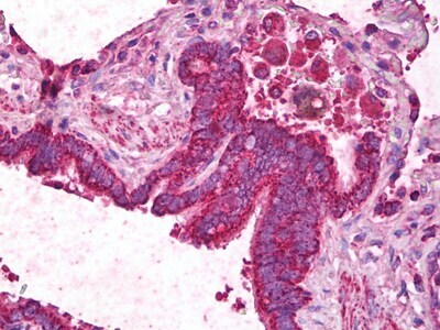
- Immunohistochemistry analysis of Angiopoietin 1 in human lung, respiratory epithelium. Samples were incubated with Angiopoietin 1 polyclonal antibody (Product # PA5-142604) using a dilution of 2.5 µg/mL. Formalin-fixed, paraffin-embedded tissue after heat-induced antigen retrieval.
Supportive validation
- Submitted by
- Invitrogen Antibodies (provider)
- Main image
- Experimental details
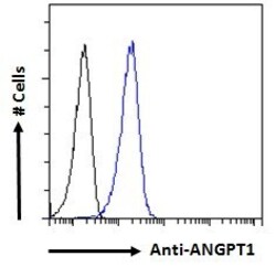
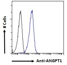
- Flow Cytometry analysis of Angiopoietin 1 in A431 cells (blue line) using Angiopoietin 1 Polyclonal Antibody (Product # PA5-142604). Cells were fixed with paraformaldehyde, permeabilized with 0.5% Triton, and incubated with the primary antibody for 1hr (10 µg/mL) followed by Alexa Fluor 488 secondary antibody (1 µg/mL). IgG control: Unimmunized goat IgG (black line) followed by Alexa Fluor 488 secondary antibody.
- Submitted by
- Invitrogen Antibodies (provider)
- Main image

- Experimental details
- Flow Cytometry analysis of Angiopoietin 1 in A431 cells (blue line) using Angiopoietin 1 Polyclonal Antibody (Product # PA5-142604). Cells were fixed with paraformaldehyde, permeabilized with 0.5% Triton, and incubated with the primary antibody for 1hr (10 µg/mL) followed by Alexa Fluor 488 secondary antibody (1 µg/mL). IgG control: Unimmunized goat IgG (black line) followed by Alexa Fluor 488 secondary antibody.
