Explore
 Validate
 Learn
Learn Western blot
 ELISA
ELISAAntibody data
- Antibody Data
- Antigen structure
- References [0]
- Comments [0]
- Validations
- Western blot [1]
- Immunohistochemistry [1]
Submit
Validation data
Reference
Comment
Report error
- Product number
- R1585 - Provider product page

- Provider
- Acris Antibodies GmbH
- Proper citation
- Acris Antibodies GmbH Cat#R1585, RRID:AB_972866
- Product name
- anti Angiopoietin-1
- Antibody type
- Polyclonal
- Antigen
- This whole rabbit serum was prepared by repeated immunizations with a synthetic peptide corresponding to a region (aa 21-40) near the N-terminus of mouse angiopoietin 1 protein conjugated to KLH using maleimide. A residue of cysteine was added to the amino terminal end to facilitate coupling.
- Reactivity
- Human, Mouse
- Host
- Rabbit
- Vial size
- 0.2 ml
- Concentration
- 85 mg/ml (by Refractometry)
No comments: Submit comment
Supportive validation
- Submitted by
- Acris Antibodies GmbH (provider)
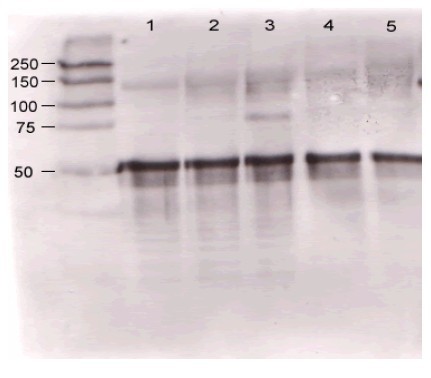
- Main image
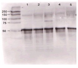
- Experimental details
- Immunoblotting. Rabbit anti-Ang 1 antibody was used at a 1/500 dilution to detect Mouse Ang-1 by western blot found in supernatants of Mouse-angiopoietin-expressing endothelial cells. Lane 1: wt endothelial cells. Lane 2: Mouse Ang-1 (clone 1-8) expressing cells. Lane 3: Mouse Ang-1 (clone 1-15) expressing cells. Lane 4: Mouse Ang-2 (clone 2-9) expressing cells.Approximately 20 µg of each lysate was loaded on a 10% SDS-PAGE. Immunoprecipitation preceded reaction with primary antibody at RT for 1 h. After subsequent washing, a 1/5,000 dilution of HRP conjugated Goat-anti Rabbit IgG (R1454HRP) preceded color development.
Supportive validation
- Submitted by
- Acris Antibodies GmbH (provider)
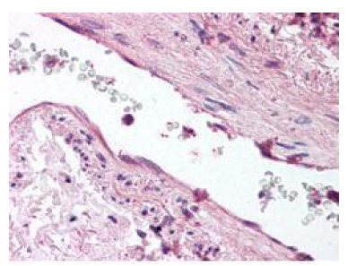
- Main image

- Experimental details
- Immunohistochemistry. Anti-ANG1 antibody was diluted 1/500 to detect ANG1 in Formalin Fixed and Paraffin Embedded Human lung tissue. Tissue was . No pre-treatment of sample was required. The image shows the localization of antibody as the precipitated red signal, with a hematoxylin purple nuclear counter stain.