Explore
 Validate
 Learn
Learn ELISA
ELISA Immunohistochemistry
Antibody data
- Antibody Data
- Antigen structure
- References [1]
- Comments [0]
- Validations
- Immunohistochemistry [1]
Submit
Validation data
Reference
Comment
Report error
- Product number
- MAB636-100 - Provider product page

- Provider
- R&D Systems
- Product name
- Human/Primate EGF Antibody
- Antibody type
- Monoclonal
- Description
- Protein A or G purified from hybridoma culture supernatant. Detects human and primate EGF in ELISAs. In sandwich ELISAs, no cross-reactivity or interference is observed with recombinant mouse (rm) EGF, bovine FGF acidic, bovine FGF basic, recombinant human (rh) G-CSF, rhGM-CSF, rmGM-CSF, rhLIF, human PDGF, porcine PDGF, rhTGF-alpha , hTGF-beta 1, pTGF-beta 1, rhTGF-beta 1, porcine TGF-beta 1.2, porcine TGF-beta 2, rhTNF-alpha , or rhTNF-beta .
- Reactivity
- Human, Simian
- Host
- Mouse
- Conjugate
- Unconjugated
- Isotype
- IgG
- Antibody clone number
- 10827
- Vial size
- 100 ug
- Storage
- Use a manual defrost freezer and avoid repeated freeze-thaw cycles. 12 months from date of receipt, -20 to -70 °C as supplied. 1 month, 2 to 8 °C under sterile conditions after reconstitution. 6 months, -20 to -70 °C under sterile conditions after reconstitution.
Submitted references Pre-analytical effects of blood sampling and handling in quantitative immunoassays for rheumatoid arthritis.
Zhao X, Qureshi F, Eastman PS, Manning WC, Alexander C, Robinson WH, Hesterberg LK
Journal of immunological methods 2012 Apr 30;378(1-2):72-80
Journal of immunological methods 2012 Apr 30;378(1-2):72-80
No comments: Submit comment
Supportive validation
- Submitted by
- R&D Systems (provider)
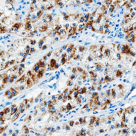
- Main image

- Experimental details
- EGF in Human Lung Cancer Tissue. EGF was detected in immersion fixed paraffin-embedded sections of human lung cancer tissue using Mouse Anti-Human/Primate EGF Monoclonal Antibody (Catalog # MAB636) at 15 µg/mL overnight at 4 °C. Tissue was stained using the Anti-Mouse HRP-DAB Cell & Tissue Staining Kit (brown; Catalog # CTS002) and counterstained with hematoxylin (blue). Specific staining was localized to the plasma membrane and cytoplasm. View our protocol for Chromogenic IHC Staining of Paraffin-embedded Tissue Sections.