Explore
 Validate
 Learn
Learn Western blot
Antibody data
- Antibody Data
- Antigen structure
- References [1]
- Comments [0]
- Validations
- Western blot [2]
- Immunohistochemistry [1]
- Other assay [1]
Submit
Validation data
Reference
Comment
Report error
- Product number
- PA5-36048 - Provider product page

- Provider
- Invitrogen Antibodies
- Product name
- Cyclin A Polyclonal Antibody
- Antibody type
- Polyclonal
- Antigen
- Synthetic peptide
- Description
- This antibody detects endogenous protein at a molecular weight of 52 kDa.
- Concentration
- 1 mg/mL
Submitted references Ablation of CRBN induces loss of type I collagen and SCH in mouse skin by fibroblast senescence via the p38 MAPK pathway.
Jeon S, Yoon YS, Kim HK, Han J, Lee KM, Seol JE, Cho SK, Park CS
Aging 2021 Mar 3;13(5):6406-6419
Aging 2021 Mar 3;13(5):6406-6419
No comments: Submit comment
Supportive validation
- Submitted by
- Invitrogen Antibodies (provider)
- Main image
- Experimental details
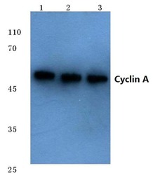
- Western blot analysis of Cyclin A using Cyclin A polyclonal antibody (Product # PA5-36048) at a dilution of 1:500. Lane 1: HEK293T cell lysate, Lane 2: NIH-3T3 cell lysate, Lane 3: Rat brain tissue lysate.

- Submitted by
- Invitrogen Antibodies (provider)
- Main image
- Experimental details
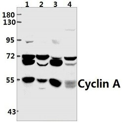
- Western blot analysis of Cyclin A in Lane 1: K562 whole cell lysate (40 µg), Lane 2: SGC7901 whole cell lysate (40 µg), Lane 3: the Testis tissue lysate of mouse (40 µg), Lane 4: the Testis tissue lysate of rat (40 µg). Samples were incubated with Cyclin A polyclonal antibody (Product # PA5-36048) at a dilution of 1:500.

Supportive validation
- Submitted by
- Invitrogen Antibodies (provider)
- Main image
- Experimental details
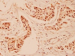
- Immunohistochemical analysis of Cyclin an In paraffin-embedded human breast carcinoma using Cyclin A polyclonal antibody (Product # PA5-36048) at a dilution of 1:100.

Supportive validation
- Submitted by
- Invitrogen Antibodies (provider)
- Main image

- Experimental details
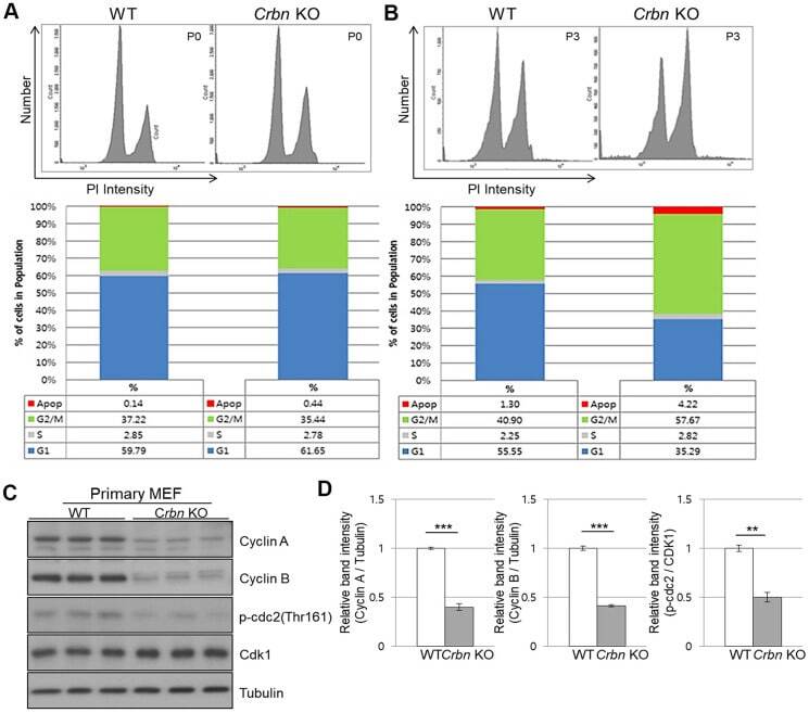
- Figure 4 CRBN deficient fibroblast exhibits G2/M cell cycle arrest. ( A , B ) PI staining and cell cycle distribution analysis of WT and CRBN KO Primary MEFs in P0 and P3. Cells were stained with PI and analyzed for cell cycle distribution using flow cytometry. Representative images of flow cytometry plots are shown. The graph indicates the distribution of each cell cycle phase with different colors, G0/G1(blue), S(gray), and G2/M(green) phases. ( C ) Western blots analysis using extracts of MEF cells in the early passage were immunoblotted with the anti-Cyclin A, anti-Cyclin B, anti-Cdk1, anti-p-cdc2, and anti-Tubulin antibodies. Tubulin was used to confirm equal protein loading. ( D ) Relative band intensities as determined by densitometric analysis of the blots in ( C ). The results shown are representative of five independent experiments. * P < 0.05; ** P < 0.01; *** P < 0.005; n.s., not significant.