 Explore
 Validate
 Learn
Learn Western blot
Antibody data
- Antibody Data
- Antigen structure
- References [1]
- Comments [0]
- Validations
- Western blot [1]
- Immunohistochemistry [1]
Submit
Validation data
Reference
Comment
Report error
- Product number
- MAB7046 - Provider product page

- Provider
- R&D Systems
- Product name
- Human Cyclin A1 Antibody
- Antibody type
- Monoclonal
- Description
- Protein A or G purified from hybridoma culture supernatant. Detects human Cyclin A1 in direct ELISAs and Western blots. In direct ELISAs and Western blots, no cross-reactivity with recombinant human Cyclin A2, B1, B2, C, D1, D2, D3, E1, E2, or recombinant mouse Cyclin A1 is observed.
- Reactivity
- Human
- Host
- Mouse
- Conjugate
- Unconjugated
- Antigen sequence
P78396- Isotype
- IgG
- Antibody clone number
- 722407
- Vial size
- 100 ug
- Concentration
- LYOPH
- Storage
- Use a manual defrost freezer and avoid repeated freeze-thaw cycles. 12 months from date of receipt, -20 to -70 °C as supplied. 1 month, 2 to 8 °C under sterile conditions after reconstitution. 6 months, -20 to -70 °C under sterile conditions after reconstitution.
Submitted references Androgen Receptor-Mediated Growth Suppression of HPr-1AR and PC3-Lenti-AR Prostate Epithelial Cells.
Kim YC, Chen C, Bolton EC
PloS one 2015;10(9):e0138286
PloS one 2015;10(9):e0138286
No comments: Submit comment
Supportive validation
- Submitted by
- R&D Systems (provider)
- Main image
- Experimental details
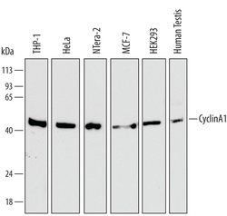
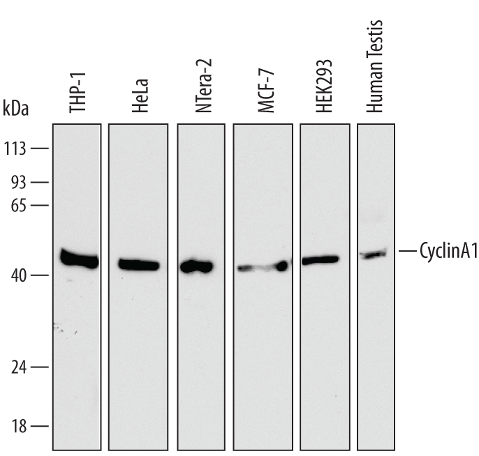
- Detection of Human Cyclin A1 by Western Blot. Western blot shows lysates of THP-1 human acute monocytic leukemia cell line, HeLa human cervical epithelial carcinoma cell line, NTera-2 human testicular embryonic carcinoma cell line, MCF-7 human breast cancer cell line, HEK293 human embryonic kidney cell line, and human testis tissue. PVDF membrane was probed with 1 µg/mL of Mouse Anti-Human Cyclin A1 Monoclonal Antibody (Catalog # MAB7046) followed by HRP-conjugated Anti-Mouse IgG Secondary Antibody (Catalog # HAF007). A specific band was detected for Cyclin A1 at approximately 52 kDa (as indicated). This experiment was conducted under reducing conditions and using Immunoblot Buffer Group 1.
Supportive validation
- Submitted by
- R&D Systems (provider)
- Main image
- Experimental details
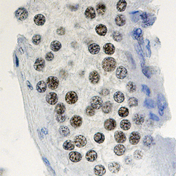
- Cyclin A1 in Human Testis. Cyclin A1 was detected in immersion fixed paraffin-embedded sections of human testis using Mouse Anti-Human Cyclin A1 Monoclonal Antibody (Catalog # MAB7046) at 15 µg/mL overnight at 4 °C. Tissue was stained using the Anti-Mouse HRP-DAB Cell & Tissue Staining Kit (brown; Catalog # CTS002) and counterstained with hematoxylin (blue). Specific staining was localized to nuclei of spermatocytes. View our protocol for Chromogenic IHC Staining of Paraffin-embedded Tissue Sections.
