Explore
 Validate
 Learn
Learn Western blot
 Immunocytochemistry
Antibody data
- Antibody Data
- Antigen structure
- References [0]
- Comments [0]
- Validations
- Western blot [2]
- Flow cytometry [2]
Submit
Validation data
Reference
Comment
Report error
- Product number
- NBP2-22632 - Provider product page

- Provider
- Novus Biologicals
- Product name
- Mouse Monoclonal Nanog Antibody
- Antibody type
- Monoclonal
- Description
- Protein A purified. It shows specificity to Nanog and is non-reactive to lysates from non-embryonal cell types (e.g. HeLa cell lysate).
- Reactivity
- Human
- Host
- Mouse
- Isotype
- IgG
- Vial size
- 100 ug
- Concentration
- 1 mg/ml
- Storage
- Store at -20C. Avoid freeze-thaw cycles.
No comments: Submit comment
Supportive validation
- Submitted by
- Novus Biologicals (provider)
- Main image

- Experimental details
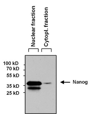
- Western Blot: Nanog Antibody (23D2-3C6) [NBP2-22632] - Analysis of 60 ug of NCCIT nuclear and cytoplasmic fractions lysates and 10ul of PageRuler Prestained Protein Ladder
- Submitted by
- Novus Biologicals (provider)
- Main image

- Experimental details
- Western Blot: Nanog Antibody (23D2-3C6) [NBP2-22632] - Analysis of 60 ug of various whole cell lysates and 10ul of PageRuler Prestained Protein Ladder.

Supportive validation
- Submitted by
- Novus Biologicals (provider)
- Main image

- Experimental details
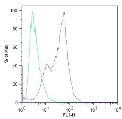
- Flow Cytometry: Nanog Antibody (23D2-3C6) [NBP2-22632] - Analysis of Nanog (blue histogram) on HEL 11.4 induced IPS cells. To generate single cells suspensions, colonies were treated with TrypLE cell dissociation enzyme for 5 minutes at 37C. Cells were incubated with a Nanog monoclonal antibody or mouse IgG (green histogram) at a dilution of 1:100 for 1 hour on ice, washed with PBS + 5% fetal calf serum (FACS buffer), and incubated with a fluorescein-conjugated secondary antibody at a dilution of 1:200 for 30 minutes on ice. Cells were washed with cold FACS buffer, resuspended in 500ul of FACS buffer containing 10ul of 4% paraformaldehyde.
- Submitted by
- Novus Biologicals (provider)
- Main image
- Experimental details
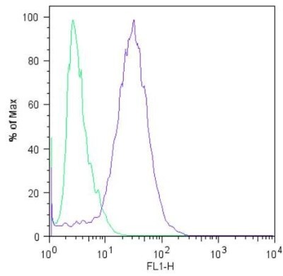
- Flow Cytometry: Nanog Antibody (23D2-3C6) [NBP2-22632] - Analysis of Nanog (blue histogram) on H9 embryonic stem cells. To generate single cells suspensions, colonies were treated with TrypLE cell dissociation enzyme for 5 minutes at 37C. Cells were incubated with a Nanog monoclonal antibody or mouse IgG (green histogram) at a dilution of 1:100 for 1 hour on ice, washed with PBS + 5% fetal calf serum (FACS buffer), and incubated with a fluorescein-conjugated secondary antibody at a dilution of 1:200 for 30 minutes on ice. Cells were washed with cold FACS buffer, resuspended in 500ul of FACS buffer containing 10ul of 4% paraformaldehyde.