Explore
 Validate
 Learn
Learn Western blot
Antibody data
- Antibody Data
- Antigen structure
- References [8]
- Comments [0]
- Validations
- Western blot [3]
- Immunocytochemistry [3]
- Flow cytometry [2]
Submit
Validation data
Reference
Comment
Report error
- Product number
- MA1-017 - Provider product page

- Provider
- Invitrogen Antibodies
- Product name
- Nanog Monoclonal Antibody (23D2-3C6)
- Antibody type
- Monoclonal
- Antigen
- Recombinant full-length protein
- Description
- Western blot analysis of MA1-017 detects an ~38 kDa protein in embryonal carcinoma cells. Subcellular fractionation shows nuclear localization of Nanog. MA1-017 shows specificity to Nanog and is non-reactive to lysates from non-embryonal cell types (e.g. HeLa cell lysate).
- Reactivity
- Human
- Host
- Mouse
- Isotype
- IgG
- Antibody clone number
- 23D2-3C6
- Vial size
- 100 µg
- Concentration
- 1 mg/mL
- Storage
- -20°C
Submitted references Buprenorphine Exposure Alters the Development and Migration of Interneurons in the Cortex.
Generation of four induced pluripotent stem cell lines (FHUi003-A, FHUi003-B, FHUi004-A and FHUi004-B) from two affected individuals of a familial neurohypophyseal diabetes insipidus family.
Fluid shear stress induces cancer stem cell-like phenotype in MCF7 breast cancer cell line without inducing epithelial to mesenchymal transition.
Generation of an iPSC line from a patient with GTP cyclohydrolase 1 (GCH1) deficiency: HDMC0061i-GCH1.
Generation of an iPSC line from a patient with tyrosine hydroxylase (TH) deficiency: TH-1 iPSC.
Conservative site-specific and single-copy transgenesis in human LINE-1 elements.
Conditionally Stabilized dCas9 Activator for Controlling Gene Expression in Human Cell Reprogramming and Differentiation.
Association of the Long Non-coding RNA Steroid Receptor RNA Activator (SRA) with TrxG and PRC2 Complexes.
Nieto-Estévez V, Donegan JJ, McMahon CL, Elam HB, Chavera TA, Varma P, Berg KA, Lodge DJ, Hsieh J
Frontiers in molecular neuroscience 2022;15:889922
Frontiers in molecular neuroscience 2022;15:889922
Generation of four induced pluripotent stem cell lines (FHUi003-A, FHUi003-B, FHUi004-A and FHUi004-B) from two affected individuals of a familial neurohypophyseal diabetes insipidus family.
Yoshida S, Okura H, Suga H, Soen M, Kawaguchi Y, Kurimoto J, Miyata T, Takagi H, Arima H, Fujikawa T, Otsuka F, Matsuyama A
Stem cell research 2020 Oct;48:101960
Stem cell research 2020 Oct;48:101960
Fluid shear stress induces cancer stem cell-like phenotype in MCF7 breast cancer cell line without inducing epithelial to mesenchymal transition.
Triantafillu UL, Park S, Klaassen NL, Raddatz AD, Kim Y
International journal of oncology 2017 Mar;50(3):993-1001
International journal of oncology 2017 Mar;50(3):993-1001
Generation of an iPSC line from a patient with GTP cyclohydrolase 1 (GCH1) deficiency: HDMC0061i-GCH1.
Jung-Klawitter S, Ebersold J, Göhring G, Blau N, Opladen T
Stem cell research 2017 Apr;20:38-41
Stem cell research 2017 Apr;20:38-41
Generation of an iPSC line from a patient with tyrosine hydroxylase (TH) deficiency: TH-1 iPSC.
Jung-Klawitter S, Blau N, Sebe A, Ebersold J, Göhring G, Opladen T
Stem cell research 2016 Nov;17(3):580-583
Stem cell research 2016 Nov;17(3):580-583
Conservative site-specific and single-copy transgenesis in human LINE-1 elements.
Vijaya Chandra SH, Makhija H, Peter S, Myint Wai CM, Li J, Zhu J, Ren Z, D'Alcontres MS, Siau JW, Chee S, Ghadessy FJ, Dröge P
Nucleic acids research 2016 Apr 7;44(6):e55
Nucleic acids research 2016 Apr 7;44(6):e55
Conditionally Stabilized dCas9 Activator for Controlling Gene Expression in Human Cell Reprogramming and Differentiation.
Balboa D, Weltner J, Eurola S, Trokovic R, Wartiovaara K, Otonkoski T
Stem cell reports 2015 Sep 8;5(3):448-59
Stem cell reports 2015 Sep 8;5(3):448-59
Association of the Long Non-coding RNA Steroid Receptor RNA Activator (SRA) with TrxG and PRC2 Complexes.
Wongtrakoongate P, Riddick G, Fucharoen S, Felsenfeld G
PLoS genetics 2015 Oct;11(10):e1005615
PLoS genetics 2015 Oct;11(10):e1005615
No comments: Submit comment
Supportive validation
- Submitted by
- Invitrogen Antibodies (provider)
- Main image
- Experimental details
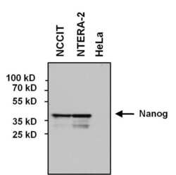
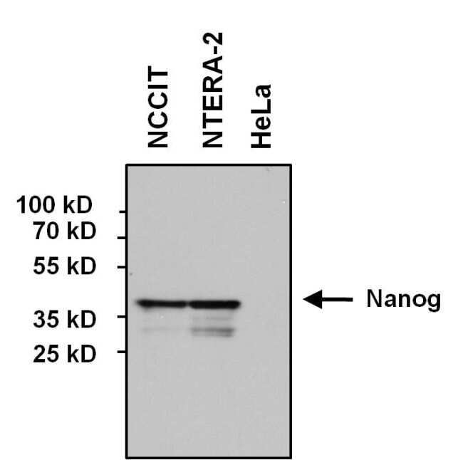
- Western blot analysis of Nanog was performed by loading 60 µg of various whole cell lysates and 10 µL of PageRuler Prestained Protein Ladder (Product # 26616) onto a 4-20% Tris-HCl polyacrylamide gel. Proteins were transferred to a PVDF membrane and blocked with 5% BSA/TBST for at least 1 hour. The membrane was probed with a Nanog monoclonal antibody (Product # MA1-017) at a dilution of 1:1000 overnight at 4°C on a rocking platform, washed in TBS-0.1%Tween-20, and probed with a goat anti-mouse IgG-HRP secondary antibody (Product # 31430) at a dilution of 1:20,000 for 1 hour. Chemiluminescent detection was performed using SuperSignal West Pico (Product # 34078). Note the absence of Nanog in negative control HeLa cell lysate.
- Submitted by
- Invitrogen Antibodies (provider)
- Main image

- Experimental details
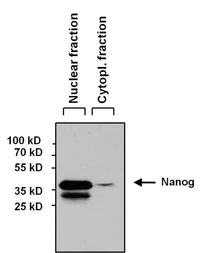
- Western blot analysis of Nanog was performed by loading 60 µg of NCCIT nuclear and cytoplasmic fractions lysates and 10 µL of PageRuler Prestained Protein Ladder (Product # 26616) onto a 4-20% Tris-HCl polyacrylamide gel. Proteins were transferred to a PVDF membrane and blocked with 5% BSA/TBST for at least 1 hour. The membrane was probed with a Nanog monoclonal antibody (Product # MA1-017) at a dilution of 1:1000 overnight at 4°C on a rocking platform, washed in TBS-0.1%Tween-20, and probed with a goat anti-mouse IgG-HRP secondary antibody (Product # 31430) at a dilution of 1:20,000 for 1 hour. Chemiluminescent detection was performed using SuperSignal West Pico (Product # 34078). Nuclear and cytoplasmic fractions were generated using the Pierce NE-PER kit (Product # 78833).
- Submitted by
- Invitrogen Antibodies (provider)
- Main image

- Experimental details
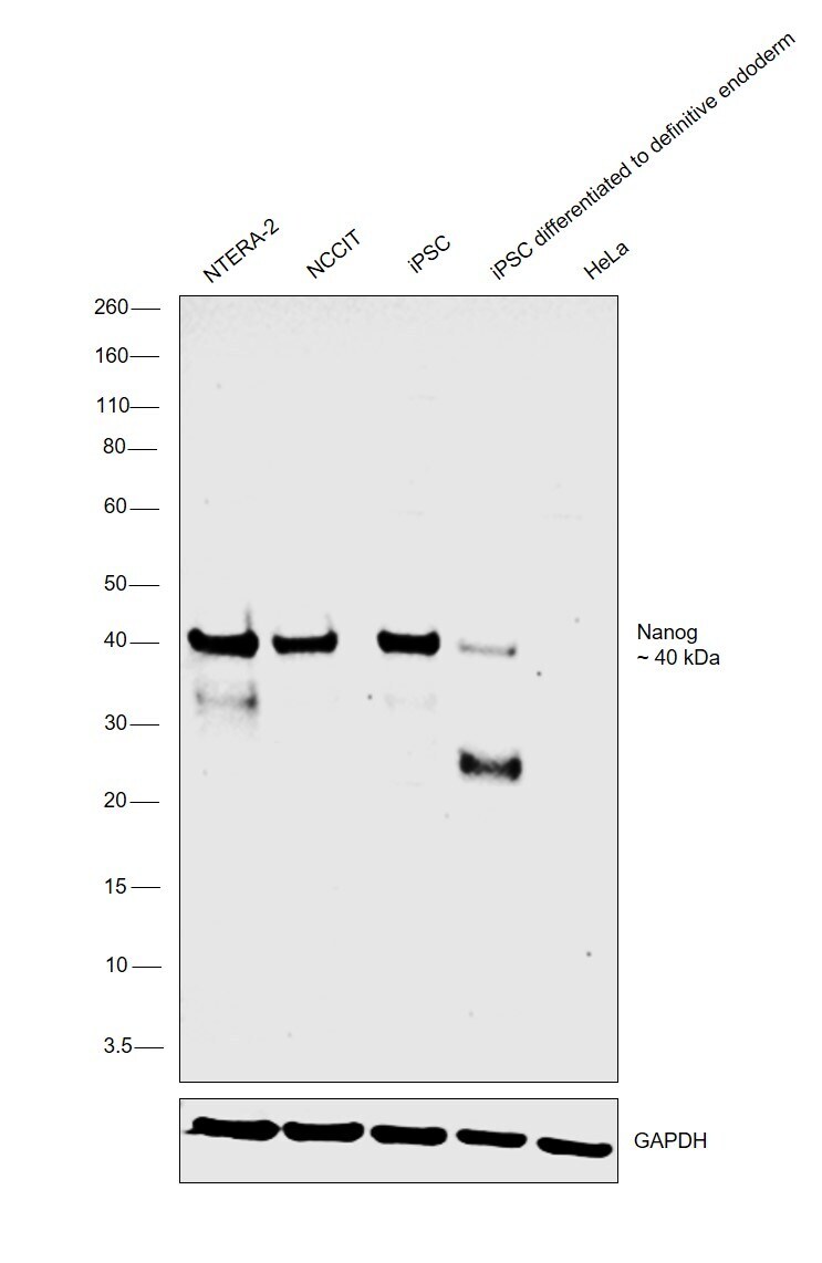
- Western blot was performed using Anti-Nanog Monoclonal Antibody (23D2-3C6) (Product # MA1-017) and a 40 kDa band corresponding to Nanog was observed across all the cell lines tested except HeLa which is reported to be negative, also the expression was decreased in iPSC upon differentiation to definitive endoderm. Modified whole cell extracts (1% SDS) (30 µg lysate) of NTERA-2 (Lane 1), NCCIT (Lane 2), iPSC (Lane 3), iPSC differentiated to definitive endoderm (Lane 4) and HeLa (Lane 5) were electrophoresed using Novex® NuPAGE® 4-12% Bis-Tris Protein Gel (Product # NP0321BOX). Resolved proteins were then transferred onto a nitrocellulose membrane (Product # IB23001) by iBlot® 2 Dry Blotting System (Product # IB21001). The blot was probed with the primary antibody (1:1000 dilution) and detected by chemiluminescence with Goat anti-Mouse IgG (H+L), Superclonal™ Recombinant Secondary Antibody, HRP (Product # A28177, 1:4000 dilution) using the iBright FL 1000 (Product # A32752). Chemiluminescent detection was performed using Novex® ECL Chemiluminescent Substrate Reagent Kit (Product # WP20005).
Supportive validation
- Submitted by
- Invitrogen Antibodies (provider)
- Main image

- Experimental details
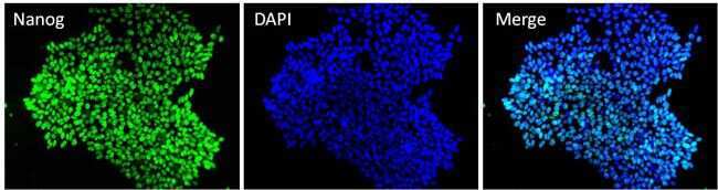
- Immunofluorescent analysis of Nanog (green) in NTERA-2 and HeLa cells. Formalin fixed cells were permeabilized with 0.1% Triton X-100 in TBS for 10 minutes at room temperature. Cells were blocked with 1% Blocker BSA (Product # 37525) for 15 minutes at room temperature. Cells were probed with a Nanog monoclonal antibody (Product # MA1-017) at a dilution of 1:50 for at least 1 hour at room temperature, washed with PBS, and incubated with a DyLight 488-conjugated goat anti-mouse IgG secondary antibody (Product # 35502). F-Actin (red) was stained with DyLight-554 Phalloidin (Product # 21834) and nuclei (blue) were stained with Hoechst 33342 dye (Product # 62249). Images were taken on a Thermo Scientific ToxInsight at 20X magnification.

- Submitted by
- Invitrogen Antibodies (provider)
- Main image

- Experimental details
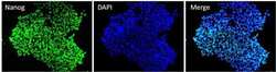
- Immunofluorescent analysis of Nanog (green) in HEL 11.4 induced IPS cells grown for a few days on Matrigel-coated chamber slides. Cells fixed in 4% paraformaldehyde were permeabilized with 0.1% Triton X-100 for 15 minutes at room temperature. Cells were probed with a Nanog monoclonal antibody (Product # MA1-017) at a dilution of 1:200 overnight at 4°C, washed with PBST, and incubated with a FITC-conjugated secondary antibody at a dilution of 1:100 for 1 hour at room temperature. Nuclei (blue) were stained with DAPI and cells were analyzed by fluorescence microscopy at 20X magnification.
- Submitted by
- Invitrogen Antibodies (provider)
- Main image
- Experimental details
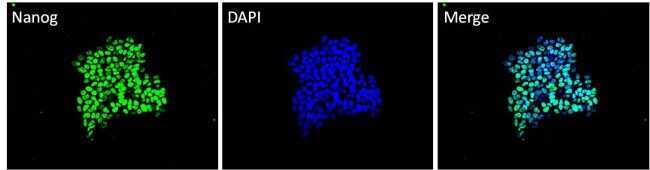
- Immunofluorescent analysis of Nanog (green) in H9 embryonic stem cells grown for a few days on Matrigel-coated chamber slides. Cells fixed in 4% paraformaldehyde were permeabilized with 0.1% Triton X-100 for 15 minutes at room temperature. Cells were probed with a Nanog monoclonal antibody (Product # MA1-017) at a dilution of 1:200 overnight at 4°C, washed with PBST, and incubated with a FITC-conjugated secondary antibody at a dilution of 1:100 for 1 hour at room temperature. Nuclei (blue) were stained with DAPI and cells were analyzed by fluorescence microscopy at 20X magnification.
Supportive validation
- Submitted by
- Invitrogen Antibodies (provider)
- Main image
- Experimental details
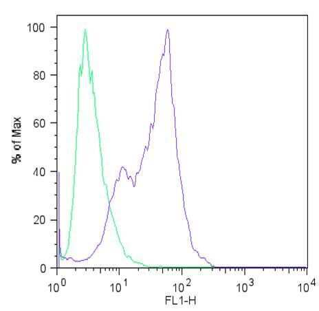
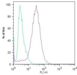
- Flow cytometric analysis of Nanog (blue histogram) on HEL 11.4 induced IPS cells. To generate single cells suspensions, colonies were treated with TrypLE cell dissociation enzyme for 5 minutes at 37°C. Cells were incubated with a Nanog monoclonal antibody (Product # MA1-017) or mouse IgG (green histogram) at a dilution of 1:100 for 1 hour on ice, washed with PBS + 5% fetal calf serum (FACS buffer), and incubated with a FITC-conjugated secondary antibody at a dilution of 1:200 for 30 minutes on ice. Cells were washed with cold FACS buffer, resuspended in 500 µL of FACS buffer containing 10 µL of 4% paraformaldehyde, and analyzed on a flow cytometer.
- Submitted by
- Invitrogen Antibodies (provider)
- Main image

- Experimental details
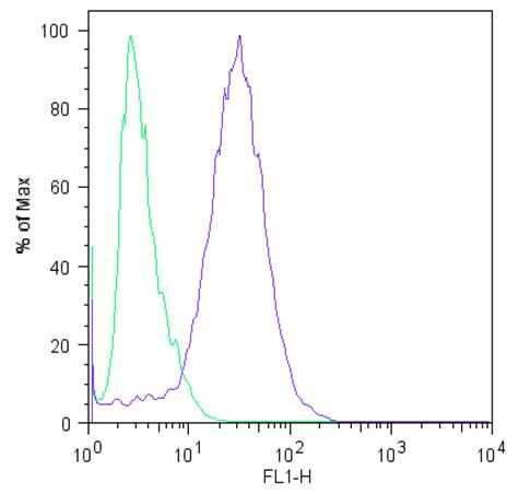
- Flow cytometric analysis of Nanog (blue histogram) on H9 embryonic stem cells. To generate single cells suspensions, colonies were treated with TrypLE cell dissociation enzyme for 5 minutes at 37°C. Cells were incubated with a Nanog monoclonal antibody (Product # MA1-017) or mouse IgG (green histogram) at a dilution of 1:100 for 1 hour on ice, washed with PBS + 5% fetal calf serum (FACS buffer), and incubated with a FITC-conjugated secondary antibody at a dilution of 1:200 for 30 minutes on ice. Cells were washed with cold FACS buffer, resuspended in 500 µL of FACS buffer containing 10 µL of 4% paraformaldehyde, and analyzed on a flow cytometer.