Explore
 Validate
 Learn
Learn Immunocytochemistry
Antibody data
- Antibody Data
- Antigen structure
- References [0]
- Comments [0]
- Validations
- Immunocytochemistry [2]
- Flow cytometry [1]
Submit
Validation data
Reference
Comment
Report error
- Product number
- MA1-017-D488 - Provider product page

- Provider
- Invitrogen Antibodies
- Product name
- Nanog Monoclonal Antibody (23D2-3C6), DyLight™ 488
- Antibody type
- Monoclonal
- Antigen
- Recombinant full-length protein
- Description
- MA1-017-D488 has been successfully used in ICC/IF applications with human samples.
- Conjugate
- Green dye
- Antibody clone number
- 23D2-3C6
- Concentration
- 1 mg/mL
No comments: Submit comment
Supportive validation
- Submitted by
- Invitrogen Antibodies (provider)
- Main image

- Experimental details
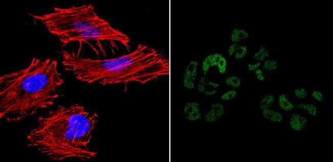
- Immunofluorescent analysis of Nanog (green) showing nuclear staining of NCCIT cells (right panel) compared to negative HeLa cell control (left panel). The cells were fixed with formalin for 15 minutes, permeabilized with 0.1% Triton X-100 in TBS, washed, and then blocked with 3% BSA-PBS for 30 minutes at room temperature. Cells were probed with a DyLight 488-conjugated Nanog monoclonal antibody (Product # MA1-017-D488) in 3% BSA-PBS at a dilution of 1:20 and incubated for 1 hour at 37C in the dark. F-Actin (left panel, red) was stained with DyLight 554 Phalloidin (Product # 21834) and nuclei (left panel, blue) were stained with DAPI. Images were taken at 60X magnification.
- Conjugate
- Green dye
- Submitted by
- Invitrogen Antibodies (provider)
- Main image

- Experimental details
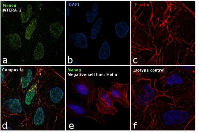
- Immunofluorescence analysis of Nanog was performed using 70% confluent log phase NTERA-2 cells. The cells were fixed with 4% paraformaldehyde for 10 minutes, permeabilized with 0.1% Triton™ X-100 for 15 minutes, and blocked with 1% BSA for 1 hour at room temperature. The cells were labeled with Nanog Mouse Monoclonal Antibody (Product # MA1-017-D488) at 1:250 dilution in 0.1% BSA, incubated at 4 degree Celsius overnight (Panel a: green). Nuclei (Panel b: blue) were stained with ProLong™ Diamond Antifade Mountant with DAPI (Product # P36962). F-actin (Panel c: red) was stained with Rhodamine Phalloidin (Product # R415, 1:300). Panel d represents the merged image showing nuclear localization. Panel e shows Nanog negative cell line HeLa with no signal. Panel f represents control cells with Isotype control to assess background. The images were captured at 60X magnification.
- Conjugate
- Green dye
Supportive validation
- Submitted by
- Invitrogen Antibodies (provider)
- Main image

- Experimental details
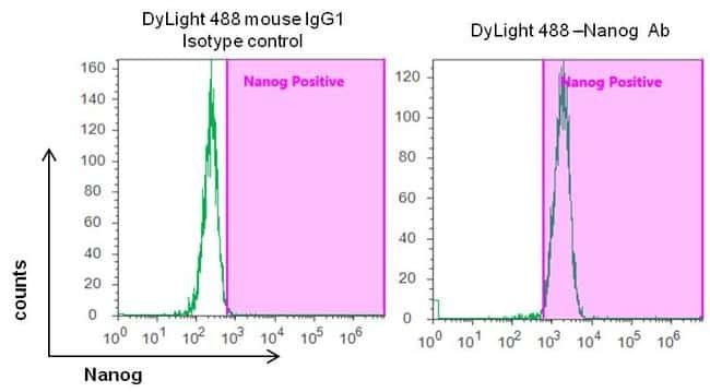
- Flow cytometry analysis of Nanog in human Gibco® Episomal iPSC line (Product # A18945) grown on vitronectin in Essential 8™ medium (Product # A1517001). Cells were fixed, permeabilized, and then stained with a 1:50 dilution of DyLight 488 directly conjugated anti-Nanog mouse monoclonal antibody (Product # MA1-017-D488) or 1:100 dilution of DyLight 488 directly conjugated mouse IgG1 isotyp control antibody (negative control, Product # MA1-191-D488) in 5% FBS/PBS. After the incubation for 1 hour on ice, the cells were analyzed on the Attune flow cytometer. A representative 10,000 cells were acquired for each sample.
- Conjugate
- Green dye