Explore
 Validate
 Learn
Learn Western blot
Antibody data
- Antibody Data
- Antigen structure
- References [0]
- Comments [0]
- Validations
- Western blot [2]
- Immunocytochemistry [1]
Submit
Validation data
Reference
Comment
Report error
- Product number
- ABIN2508894 - Provider product page

- Provider
- antibodies-online
- Product name
- anti-Nanog Homeobox (NANOG) antibody (Biotin)
- Antibody type
- Polyclonal
- Antigen
- Other
- Description
- Produced from sera of rabbits pre-immunized with highly pure (>98%) recombinant hNanog. Anti-Human Nanog specific antibody was purified by affinity chromatography and then biotinylated.
- Reactivity
- Human
- Host
- Rabbit
- Conjugate
- Biotin
- Vial size
- 50 μg
- Storage
- -20°C
No comments: Submit comment
Supportive validation
- Submitted by
- antibodies-online (provider)
- Main image

- Experimental details
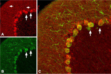
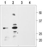
- Western blot analysis of rat glioma C6 (1, 3) or human neuroblastoma SH-SY5Y (2, 4) cell lysate: 1, 2. Anti-proBDNF antibody (ABIN2511049), (1:200). 3, 4. Anti-proBDNF antibody, preincubated with the control peptide antigen. Western blot analysis using Anti-proBDNF antibody (ABIN2511049), (1:400): 1. proBDNF (WT-human) (B-257), (20 ng). 2. proNGF (WT-human) (N-280), (200 ng). 3. recombinant proNT-3 (200 ng). 4. hBDNF (B-250), (200 ng). 5. mNGF 2.5S (Grade II) (N-100), (200 ng). 6. hNT-3 (N-260), (200 ng). Expression of proBDNF in rat cerebellum Immunohistochemical staining of rat cerebellum with Anti-proBDNF antibody (ABIN2511049). proBDNF (red) appears in Purkinje cell bodies (vertical arrows) and in astrocytic processes (horizontal arrows in A) but not in Purkinje neuronal dendrites stained for calbindin D28k (green, in B) in the same brain section. C. Confocal merge of proBDNF and CBD28K demonstrates the restriction of proBDNF to Purkinje cell body.

- Submitted by
- antibodies-online (provider)
- Main image

- Experimental details
- Western blot analysis of rat glioma C6 (1, 3) or human neuroblastoma SH-SY5Y (2, 4) cell lysate: 1, 2. Anti-proBDNF antibody (ABIN2511049), (1:200). 3, 4. Anti-proBDNF antibody, preincubated with the control peptide antigen. Western blot analysis using Anti-proBDNF antibody (ABIN2511049), (1:400): 1. proBDNF (WT-human) (B-257), (20 ng). 2. proNGF (WT-human) (N-280), (200 ng). 3. recombinant proNT-3 (200 ng). 4. hBDNF (B-250), (200 ng). 5. mNGF 2.5S (Grade II) (N-100), (200 ng). 6. hNT-3 (N-260), (200 ng). Expression of proBDNF in rat cerebellum Immunohistochemical staining of rat cerebellum with Anti-proBDNF antibody (ABIN2511049). proBDNF (red) appears in Purkinje cell bodies (vertical arrows) and in astrocytic processes (horizontal arrows in A) but not in Purkinje neuronal dendrites stained for calbindin D28k (green, in B) in the same brain section. C. Confocal merge of proBDNF and CBD28K demonstrates the restriction of proBDNF to Purkinje cell body.
Supportive validation
- Submitted by
- antibodies-online (provider)
- Main image

- Experimental details
- Image(s): Immunofluorescence