Explore
 Validate
 Learn
Learn Western blot
 ELISA
ELISAAntibody data
- Antibody Data
- Antigen structure
- References [0]
- Comments [0]
- Validations
- Western blot [2]
- Immunocytochemistry [2]
- Immunohistochemistry [1]
- Flow cytometry [1]
Submit
Validation data
Reference
Comment
Report error
- Product number
- F44247 - Provider product page

- Provider
- NSJ Bioreagents
- Product name
- NANOG Antibody (N-Terminal Region)
- Antibody type
- Polyclonal
- Description
- This highly specific NANOG antibody is suitable for use in Western blot/Immunofluorescence/Immunohistochemistry/Flow cytometry/ELISA applications with human samples.
- Reactivity
- Human
- Host
- Rabbit
- Conjugate
- Unconjugated
- Vial size
- 0.08 ml, 0.4 ml
- Concentration
- In 1X PBS, pH 7.4, with 0.09% sodium azide
- Storage
- Aliquot the NANOG antibody and store frozen at -20oC or colder. Avoid repeated freeze-thaw cycles.
No comments: Submit comment
Supportive validation
- Submitted by
- NSJ Bioreagents (provider)
- Main image
- Experimental details
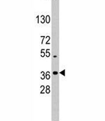
- Western blot analysis of NANOG antibody and K562 lysate; Predicted molecular weight: 35-45 kDa.

- Submitted by
- NSJ Bioreagents (provider)
- Main image
- Experimental details
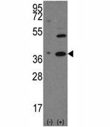
- Western blot analysis of NANOG antibody and 293 cell lysate (2 ug/lane) either nontransfected (Lane 1) or transiently transfected with the human gene (2). Predicted molecular weight: 35-45 kDa.

Supportive validation
- Submitted by
- NSJ Bioreagents (provider)
- Main image
- Experimental details
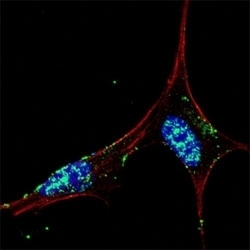
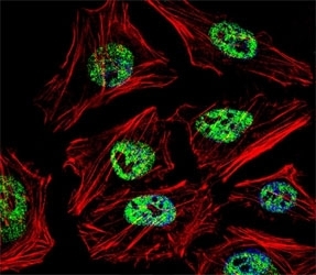
- Fluorescent confocal image of SY5Y cells stained with NANOG antibody at 1:50. Immunoreactivity is localized mainly to the nuclei of the cells.

- Submitted by
- NSJ Bioreagents (provider)
- Main image

- Experimental details
- Fluorescent confocal image of HeLa cell stained with NANOG antibody at 1:25. Immunoreactivity is localized to the nucleus.
Supportive validation
- Submitted by
- NSJ Bioreagents (provider)
- Main image

- Experimental details
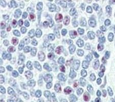
- IHC analysis of FFPE human spleen tissue stained with NANOG antibody
Supportive validation
- Submitted by
- NSJ Bioreagents (provider)
- Main image

- Experimental details
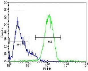
- NANOG antibody flow cytometric analysis of HepG2 cells (right histogram) compared to a negative control (left histogram). FITC-conjugated goat-anti-rabbit secondary Ab was used for the analysis.