 Explore
 Validate
 Learn
Learn Western blot
 Immunohistochemistry
Antibody data
- Antibody Data
- Antigen structure
- References [3]
- Comments [0]
- Validations
- Western blot [1]
- Immunocytochemistry [2]
- Other assay [4]
Submit
Validation data
Reference
Comment
Report error
- Product number
- PA5-18406 - Provider product page

- Provider
- Invitrogen Antibodies
- Product name
- Nanog Polyclonal Antibody
- Antibody type
- Polyclonal
- Antigen
- Synthetic peptide
- Description
- This antibody is predicted to react with canine based on sequence homology. This antibody has been successfully tested in a Sandwhich ELISA as both a capture and a detection antibody in combination with PA5-18678.
- Reactivity
- Human
- Host
- Goat
- Isotype
- IgG
- Vial size
- 100 µg
- Concentration
- 0.5 mg/mL
- Storage
- -20° C, Avoid Freeze/Thaw Cycles
Submitted references Optimization of Thymidine Kinase-Based Safety Switch for Neural Cell Therapy.
NANOG overexpression and its correlation with stem cell and differentiation markers in meningiomas of different WHO grades.
Multipotent progenitor cells derived from adult peripheral blood of swine have high neurogenic potential in vitro.
Locatelli M, Delhaes F, Cherpin O, Black ME, Carnesecchi S, Preynat-Seauve O, Hibaoui Y, Krause KH
Cells 2022 Jan 31;11(3)
Cells 2022 Jan 31;11(3)
NANOG overexpression and its correlation with stem cell and differentiation markers in meningiomas of different WHO grades.
Freitag D, McLean AL, Simon M, Koch A, Grube S, Walter J, Kalff R, Ewald C
Molecular carcinogenesis 2017 Aug;56(8):1953-1964
Molecular carcinogenesis 2017 Aug;56(8):1953-1964
Multipotent progenitor cells derived from adult peripheral blood of swine have high neurogenic potential in vitro.
Spitzer N, Sammons GS, Butts HM, Grover LM, Price EM
Journal of cellular physiology 2011 Dec;226(12):3156-68
Journal of cellular physiology 2011 Dec;226(12):3156-68
No comments: Submit comment
Supportive validation
- Submitted by
- Invitrogen Antibodies (provider)
- Main image

- Experimental details
- Western Blot staining of Human Ovary lysate using Product # PA5-18406 at a concentration of 0.03 µg/mL, the primary antibody incubation was 1 hour and the detection method was chemiluminescence.

Supportive validation
- Submitted by
- Invitrogen Antibodies (provider)
- Main image

- Experimental details
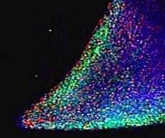
- Immunofluorescent staining (green) parts of a colony of induced pluriform stem cells derived from Human Keratinocytes using Product # PA5-18406 at a concentration of 5 µg/mL.
- Submitted by
- Invitrogen Antibodies (provider)
- Main image

- Experimental details
- Immunofluorescent staining (green) parts of a colony of induced pluriform stem cells derived from Human Keratinocytes using Product # PA5-18406 at a concentration of 5 µg/mL.

Supportive validation
- Submitted by
- Invitrogen Antibodies (provider)
- Main image

- Experimental details
- Co-localization analysis depicting the relative counts of Nanog+ cells after triple-staining for SOX2/OCT4 (A), SOX2/KI-67 (B) and Nestin/KI-67 (C). (D-I) Examples of the immunofluorescence staining (400-fold) for NANOG/SOX2/OCT4 (D + E), NANOG/SOX2/KI-67 (F + G), and NANOG/Nestin/KI-67 (H + I) for one low-grade (D + F + H) and one high-grade (E + G + I) sample

- Submitted by
- Invitrogen Antibodies (provider)
- Main image

- Experimental details
- Co-localization analysis. Specimens were examined by confocal microscopy, counting all NANOG+ cells in the tissue slide (A) and the co-labeled specific marker (B). Depicted here are the relative cell counts after double-staining. (C-F) Examples of the immunofluorescence staining (400-fold) of NANOG+ cells and nuclei (C + D), as well as NANOG and CD44 co-staining (E + F) for one low-grade (C + E) and one high-grade (E + F) sample

- Submitted by
- Invitrogen Antibodies (provider)
- Main image

- Experimental details
- Co-localization analysis depicting the relative counts of Nanog+ cells after triple-staining for SOX2/OCT4 (A), SOX2/KI-67 (B) and Nestin/KI-67 (C). (D-I) Examples of the immunofluorescence staining (400-fold) for NANOG/SOX2/OCT4 (D + E), NANOG/SOX2/KI-67 (F + G), and NANOG/Nestin/KI-67 (H + I) for one low-grade (D + F + H) and one high-grade (E + G + I) sample

- Submitted by
- Invitrogen Antibodies (provider)
- Main image
- Experimental details
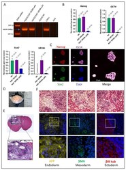
- Figure 2 Characterization of the SR39h-expressing hESC line. ( A ) PCR analysis of SR39h transgene insertion in genomic DNA. ( B ) RT-PCR analysis of the expression of pluripotency genes ( n = 3), OCT-4, NANOG, SOX2 , and SR39h in SR39h-expressing hESC (undifferentiated and differentiated), as well as control wild type cells (undifferentiated). ( C ) Immunofluorescence staining of pluripotency markers (OCT4, SOX2, NANOG), nuclei were counterstained with Hoechst. The merge panel consists of all four staining. Scale bar 20 uM. ( D ) Macroscopically visible teratoma (blue circle) after brain extraction from the skull bone. ( E ) Hematoxylin and eosin staining of coronal slice of mice brain. ( F ) Upper panel; coronal sections brain with teratoma stained with hematoxylin and eosin. Scale bar 100 uM. Lower panels; immunostaining: alpha-fetoprotein (AFP) for endoderm-derived tissue, alpha-smooth muscle actin (SMA) for mesoderm-derived tissue and Beta-III tubulin (betaIII tub) for ectoderm-derived tissue.
