 Explore
 Validate
 Learn
Learn Western blot
Antibody data
- Antibody Data
- Antigen structure
- References [3]
- Comments [0]
- Validations
- Western blot [3]
- Immunocytochemistry [3]
- Immunohistochemistry [1]
- Other assay [1]
Submit
Validation data
Reference
Comment
Report error
- Product number
- PA5-20889 - Provider product page

- Provider
- Invitrogen Antibodies
- Product name
- Nanog Polyclonal Antibody
- Antibody type
- Polyclonal
- Antigen
- Synthetic peptide
- Description
- A suggested positive control is human spleen tissue lysate. PA5-20889 can be used with blocking peptide PEP-1003.
- Reactivity
- Human, Mouse, Rat
- Host
- Rabbit
- Isotype
- IgG
- Vial size
- 100 µg
- Concentration
- 1 mg/mL
- Storage
- Maintain refrigerated at 2-8°C for up to 3 months. For long term storage store at -20°C
Submitted references Primo Vascular Node in the Bone Marrow and Longevity.
Hemmule: A Novel Structure with the Properties of the Stem Cell Niche.
An Effective and Reliable Xeno-free Cryopreservation Protocol for Single Human Pluripotent Stem Cells.
Vodyanoy V, Pustovyy O, Globa L
Journal of acupuncture and meridian studies 2022 Feb 28;15(1):12-24
Journal of acupuncture and meridian studies 2022 Feb 28;15(1):12-24
Hemmule: A Novel Structure with the Properties of the Stem Cell Niche.
Vodyanoy V, Pustovyy O, Globa L, Kulesza RJ Jr, Sorokulova I
International journal of molecular sciences 2020 Jan 14;21(2)
International journal of molecular sciences 2020 Jan 14;21(2)
An Effective and Reliable Xeno-free Cryopreservation Protocol for Single Human Pluripotent Stem Cells.
Meng G, Poon A, Liu S, Rancourt DE
Methods in molecular biology (Clifton, N.J.) 2016;1516:47-56
Methods in molecular biology (Clifton, N.J.) 2016;1516:47-56
No comments: Submit comment
Supportive validation
- Submitted by
- Invitrogen Antibodies (provider)
- Main image

- Experimental details
- Western blot analysis of human spleen tissue lysate using a NANOG polyclonal antibody (Product # PA5-20889) at (A) 1 and (B) 2 µg/mL.

- Submitted by
- Invitrogen Antibodies (provider)
- Main image

- Experimental details
- Western Blot analysis of NANOG in human spleen tissue lysate with Nanog Polyclonal Antibody (Product # PA5-20889) at (A) 1 and (B) 2 µg/mL.

- Submitted by
- Invitrogen Antibodies (provider)
- Main image

- Experimental details
- Western blot was performed using Anti-Nanog Polyclonal Antibody (Product # PA5-20889) and a band at 40 kDa corresponding to Nanog was observed across the cell lines and tissues tested along with an uncharacterized band (*) at ~ 50 kDa in the cell lines. Modified whole cell extracts (1% SDS) (30 µg lysate) of F9 (Lane 1), NIH/3T3 (Lane 2), C2C12 (Lane 3), Neuro-2a (Lane 4), Mouse Spleen (Lane 5), Rat Spleen (Lane 6) Mouse Liver (Lane 7) were electrophoresed using Novex® NuPAGE® 4-12% % Bis-Tris gel (Product # NP0321BOX). Resolved proteins were then transferred onto a nitrocellulose membrane (Product # IB23001) by iBlot® 2 Dry Blotting System (Product # IB21001). The blot was probed with the primary antibody (2 µg/mL) and detected by chemiluminescence with Goat anti-Rabbit IgG (H+L) Superclonal™ Recombinant Secondary Antibody, HRP (Product # A27036, 1:4000 dilution) using the iBright FL 1000 (Product # A32752). Chemiluminescent detection was performed using Novex® ECL Chemiluminescent Substrate Reagent Kit (Product # WP20005).

Supportive validation
- Submitted by
- Invitrogen Antibodies (provider)
- Main image
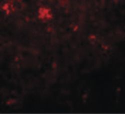
- Experimental details
- Immunofluorescent analysis of human spleen cells using a NANOG polyclonal antibody (Product # PA5-20889) at a 20 µg/mL dilution.

- Submitted by
- Invitrogen Antibodies (provider)
- Main image
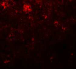
- Experimental details
- Immunofluorescence of NANOG in Human Spleen cells with Nanog Polyclonal Antibody (Product # PA5-20889) at 20 µg/mL.

- Submitted by
- Invitrogen Antibodies (provider)
- Main image
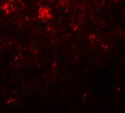
- Experimental details
- Immunofluorescence of NANOG in Human Spleen cells with Nanog Polyclonal Antibody (Product # PA5-20889) at 20 µg/mL.

Supportive validation
- Submitted by
- Invitrogen Antibodies (provider)
- Main image
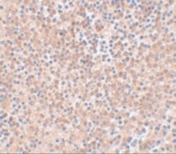
- Experimental details
- Immunohistochemistry of NANOG in human spleen tissue with Nanog Polyclonal Antibody (Product # PA5-20889) at 5 µg/mL.

Supportive validation
- Submitted by
- Invitrogen Antibodies (provider)
- Main image

- Experimental details
- Figure 7 Individual hemmule cells stained by stem cell antibodies. ( A ) Fluorescent images of individual stem cell progenitors in the hemmule, which express embryonic cell markers. The top sections show individual stem cell progenitors being scattered in the hemmule cross-sections. The lower sections denote the progenitor cells inside the hemmule vessels L--lumen. Bars in the top panels, Oct4, 50 mum; Nanog, 20 mum; CD150, 10 mum; CD90, 20 mum; CD133, 10 mum. Bars in lower panels: Oct4, 20 mum; Nanog, 20 mum; CD150, 10 mum; CD90, 10 mum; CD133, 10 mum. ( B ) Large size cells in the hemmule belong to the megakaryocyte family. Some of these large sized cells illustrate small cytoplasmic extension (depicted by red arrowhead) and are in close proximity to the pores--the optically empty rounded regions (arrows). The formation of the most of these pores occurs when the large sized cells are displaced during the thin histological sample slicing. The rims of these pores may help retain molecules from these cells, which is evidenced by the staining by the antibodies. Bars: 10 mum. ( C ) Borders of the pores are stained by antibodies. ( D ) Both bone marrow and blood vessel cells stained by stem cell antibodies (positive controls). ( a ) Anti-CD133 positive cells in the bone marrow: the large size cell (arrow) and scattered cells (arrowheads). ( b ) Anti-CD133 positive scattered cells in bone marrow. ( c ) Anti-OCT4 antibody positive cells (arrow) on the surface of the blood vessel. (
