Explore
 Validate
 Learn
Learn Western blot
Antibody data
- Antibody Data
- Antigen structure
- References [2]
- Comments [0]
- Validations
- Western blot [1]
- Immunocytochemistry [2]
- Other assay [1]
Submit
Validation data
Reference
Comment
Report error
- Product number
- PA5-11856 - Provider product page

- Provider
- Invitrogen Antibodies
- Product name
- BMPR1A Polyclonal Antibody
- Antibody type
- Polyclonal
- Antigen
- Synthetic peptide
- Reactivity
- Human
- Host
- Rabbit
- Isotype
- IgG
- Vial size
- 400 μL
- Concentration
- 2 mg/mL
- Storage
- Store at 4°C short term. For long term storage, store at -20°C, avoiding freeze/thaw cycles.
Submitted references Nanovibrational Stimulation of Mesenchymal Stem Cells Induces Therapeutic Reactive Oxygen Species and Inflammation for Three-Dimensional Bone Tissue Engineering.
Material-driven fibronectin assembly for high-efficiency presentation of growth factors.
Orapiriyakul W, Tsimbouri MP, Childs P, Campsie P, Wells J, Fernandez-Yague MA, Burgess K, Tanner KE, Tassieri M, Meek D, Vassalli M, Biggs MJP, Salmeron-Sanchez M, Oreffo ROC, Reid S, Dalby MJ
ACS nano 2020 Aug 25;14(8):10027-10044
ACS nano 2020 Aug 25;14(8):10027-10044
Material-driven fibronectin assembly for high-efficiency presentation of growth factors.
Llopis-Hernández V, Cantini M, González-García C, Cheng ZA, Yang J, Tsimbouri PM, García AJ, Dalby MJ, Salmerón-Sánchez M
Science advances 2016 Aug;2(8):e1600188
Science advances 2016 Aug;2(8):e1600188
No comments: Submit comment
Supportive validation
- Submitted by
- Invitrogen Antibodies (provider)
- Main image
- Experimental details
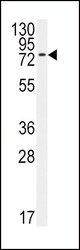
- Western blot analysis of BMPR1A in 293 cell line lysates. Samples were incubated with BMPR1A polyclonal antibody (Product # PA5-11856). Lysates: 35 µg/lane. BMPR1A (arrow).
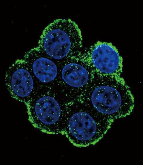
Supportive validation
- Submitted by
- Invitrogen Antibodies (provider)
- Main image
- Experimental details
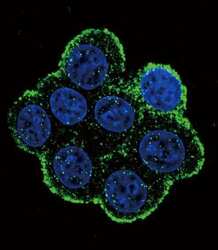
- Immunofluorescent analysis of 293 cells using a BMPR1A polyclonal antibody (Product # PA5-11856) at a dilution of 1:10-50, followed by a fluor-conjugated goat anti-rabbit secondary antibody (green). Nuclei were stained with DAPI (blue).
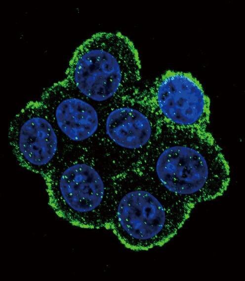
- Submitted by
- Invitrogen Antibodies (provider)
- Main image
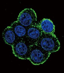
- Experimental details
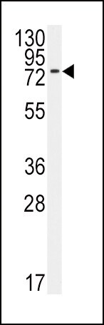
- Immunocytochemistry analysis of BMPR1A in 293 cells. Samples were incubated in BMPR1A polyclonal antibody (Product # PA5-11856) followed by Alexa Fluor 488-conjugated goat anti-rabbit lgG (green). DAPI was used to stain the cell nuclear (blue).
Supportive validation
- Submitted by
- Invitrogen Antibodies (provider)
- Main image

- Experimental details
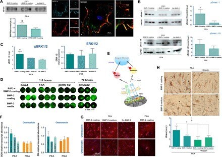
- Fig. 2 Integrin/BMP-2 receptor cosignaling drives MSC osteogenesis. ( A ) Coimmunoprecipitation of integrin beta 1 and BMPRI occurred on BMP-2 sequestered by FN on PEA, and bands correspond to BMPRIa (60 kD) after precipitation with anti-integrin beta 1 antibodies. The graphs show quantification of bands relative to the absence of BMP-2. This colocalization can also be seen in individual cells with integrin beta 1 (stained red) and BMPRIa (stained green). ( B ) Smad signaling was drastically altered when BMP-2 was presented bound on FNIII 12-14 ; blocking this GF-binding domain of FN (using the monoclonal antibody P5F3 at a molar ratio of 1 with FN to block the GF-binding site) reduces Smad signaling. GAPDH, glyceraldehyde-3-phosphate dehydrogenase. ( C ) Phosphorylation of extracellular signal-related kinase (ERK) 1/2 was significantly enhanced on PEA when BMP-2 was presented at the material interface, sequestered on FN, compared to the presence of the same doses of the soluble factor. ( D ) In-cell Western assay for Smad, FAK, pERK 1/2, and pRUNX2 with BMP-2 on FN on PEA, soluble BMP-2, and blocking with P5F3 before BMP-2 adsorption. ( E ) This fulfills the first part of the synergistic signaling hypothesis. ( F ) Quantitative polymerase chain reaction (qPCR) for osteocalcin (OCN) and osteonectin (ON) after 14 days of culture (PEA and PMA); enhanced expression occurs when BMP-2 was presented bound on FN compared to soluble administration of the GF or when BMP-2 was sequeste