 Explore
 Validate
 Learn
Learn Western blot
Antibody data
- Antibody Data
- Antigen structure
- References [1]
- Comments [0]
- Validations
- Western blot [2]
- Immunohistochemistry [1]
Submit
Validation data
Reference
Comment
Report error
- Product number
- AF5134 - Provider product page

- Provider
- R&D Systems
- Product name
- Human/Mouse/Rat PKC epsilon Antibody
- Antibody type
- Polyclonal
- Description
- Antigen Affinity-purified. Detects human, mouse and rat PKC epsilon in Western blots.
- Reactivity
- Human, Mouse, Rat
- Host
- Sheep
- Conjugate
- Unconjugated
- Antigen sequence
Q02156- Isotype
- IgG
- Vial size
- 100 ug
- Concentration
- LYOPH
- Storage
- Use a manual defrost freezer and avoid repeated freeze-thaw cycles. 12 months from date of receipt, -20 to -70 °C as supplied. 1 month, 2 to 8 °C under sterile conditions after reconstitution. 6 months, -20 to -70 °C under sterile conditions after reconstitution.
Submitted references Quercetin ameliorates paclitaxel-induced neuropathic pain by stabilizing mast cells, and subsequently blocking PKCε-dependent activation of TRPV1.
Gao W, Zan Y, Wang ZJ, Hu XY, Huang F
Acta pharmacologica Sinica 2016 Sep;37(9):1166-77
Acta pharmacologica Sinica 2016 Sep;37(9):1166-77
No comments: Submit comment
Supportive validation
- Submitted by
- R&D Systems (provider)
- Main image
- Experimental details
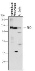
- Detection of Human/Mouse/Rat PKC epsilon by Western Blot. Western blot shows lysates of human, mouse, and rat brain tissue. PVDF membrane was probed with 1 µg/mL of Human/Mouse/Rat PKC epsilon Antigen Affinity-purified Polyclonal Antibody (Catalog # AF5134) followed by HRP-conjugated Anti-Sheep IgG Secondary Antibody (Catalog # HAF016). A specific band was detected for PKC epsilon at approximately 90 kDa (as indicated). This experiment was conducted under reducing conditions and using Immunoblot Buffer Group 1.
- Submitted by
- R&D Systems (provider)
- Main image

- Experimental details
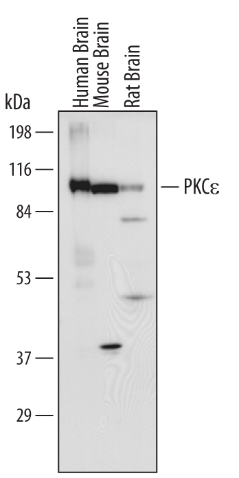
- Detection of Human PKC epsilon by Western Blot. Western blot shows lysates of human brain (hippocampus) tissue and human brain (hypothalamus) tissue. PVDF membrane was probed with 1 µg/mL of Sheep Anti-Human/Mouse/Rat PKC epsilon Antigen Affinity-purified Polyclonal Antibody (Catalog # AF5134) followed by HRP-conjugated Anti-Sheep IgG Secondary Antibody (Catalog # HAF016). A specific band was detected for PKC epsilon at approximately 90 kDa (as indicated). This experiment was conducted under reducing conditions and using Immunoblot Buffer Group 1.

Supportive validation
- Submitted by
- R&D Systems (provider)
- Main image
- Experimental details
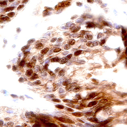
- PKC epsilon in Human Lung Cancer Tissue. PKC epsilon was detected in immersion fixed paraffin-embedded sections of human lung cancer tissue using Human/Mouse/Rat PKC epsilon Antigen Affinity-purified Polyclonal Antibody (Catalog # AF5134) at 15 µg/mL overnight at 4 °C. Before incubation with the primary antibody, tissue was subjected to heat-induced epitope retrieval using Antigen Retrieval Reagent-Basic (Catalog # CTS013). Tissue was stained using the Anti-Sheep HRP-DAB Cell & Tissue Staining Kit (brown; Catalog # CTS019) and counterstained with hematoxylin (blue). Specific staining was localized to nuclei. View our protocol for Chromogenic IHC Staining of Paraffin-embedded Tissue Sections.
