Explore
 Validate
 Learn
Learn Western blot
 Immunohistochemistry
Antibody data
- Antibody Data
- Antigen structure
- References [5]
- Comments [0]
- Validations
- Immunohistochemistry [1]
Submit
Validation data
Reference
Comment
Report error
- Product number
- HPA020896 - Provider product page

- Provider
- Atlas Antibodies
- Proper citation
- Atlas Antibodies Cat#HPA020896, RRID:AB_1845117
- Product name
- Anti-ASS1
- Antibody type
- Polyclonal
- Description
- Polyclonal Antibody against Human ASS1, Gene description: argininosuccinate synthase 1, Alternative Gene Names: ASS, CTLN1, Validated applications: IHC, WB, Uniprot ID: P00966, Storage: Store at +4°C for short term storage. Long time storage is recommended at -20°C.
- Reactivity
- Human
- Host
- Rabbit
- Conjugate
- Unconjugated
- Isotype
- IgG
- Vial size
- 100 µl
- Concentration
- 0.1 mg/ml
- Storage
- Store at +4°C for short term storage. Long time storage is recommended at -20°C.
- Handling
- The antibody solution should be gently mixed before use.
Submitted references Pancreatic tumors exhibit myeloid-driven amino acid stress and upregulate arginine biosynthesis
Cationic Amino Acid Transporter-1-Mediated Arginine Uptake Is Essential for Chronic Lymphocytic Leukemia Cell Proliferation and Viability
Argininosuccinate Synthase 1-Deficiency Enhances the Cell Sensitivity to Arginine through Decreased DEPTOR Expression in Endometrial Cancer
Gene‐specific correlation of RNA and protein levels in human cells and tissues
Proteomic analysis of oropharyngeal carcinomas reveals novel HPV-associated biological pathways.
Dzierozynski L, Apiz Saab J, Jonker P, AminiTabrizi R, Shah H, Menjivar R, Scott A, Nwosu Z, Zhu Z, Chen R, Oh M, Sheehan C, Wahl D, Pasca di Magliano M, Lyssiotis C, Macleod K, Weber C, Muir A
eLife 2023;12
eLife 2023;12
Cationic Amino Acid Transporter-1-Mediated Arginine Uptake Is Essential for Chronic Lymphocytic Leukemia Cell Proliferation and Viability
Werner A, Pieh D, Echchannaoui H, Rupp J, Rajalingam K, Theobald M, Closs E, Munder M
Frontiers in Oncology 2019;9
Frontiers in Oncology 2019;9
Argininosuccinate Synthase 1-Deficiency Enhances the Cell Sensitivity to Arginine through Decreased DEPTOR Expression in Endometrial Cancer
Ohshima K, Nojima S, Tahara S, Kurashige M, Hori Y, Hagiwara K, Okuzaki D, Oki S, Wada N, Ikeda J, Kanai Y, Morii E
Scientific Reports 2017;7(1)
Scientific Reports 2017;7(1)
Gene‐specific correlation of RNA and protein levels in human cells and tissues
Edfors F, Danielsson F, Hallström B, Käll L, Lundberg E, Pontén F, Forsström B, Uhlén M
Molecular Systems Biology 2016;12(10)
Molecular Systems Biology 2016;12(10)
Proteomic analysis of oropharyngeal carcinomas reveals novel HPV-associated biological pathways.
Slebos RJ, Jehmlich N, Brown B, Yin Z, Chung CH, Yarbrough WG, Liebler DC
International journal of cancer 2013 Feb 1;132(3):568-79
International journal of cancer 2013 Feb 1;132(3):568-79
No comments: Submit comment
Supportive validation
- Submitted by
- Atlas Antibodies (provider)
- Enhanced method
- Orthogonal validation
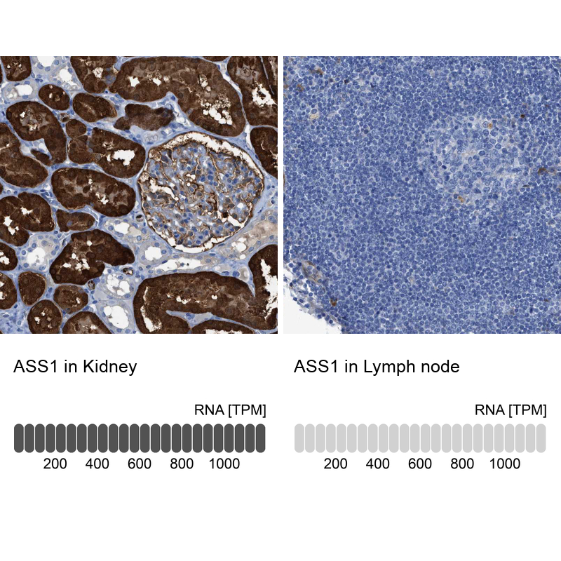
- Main image
- Experimental details
- Immunohistochemistry analysis in human kidney and lymph node tissues using HPA020896 antibody. Corresponding ASS1 RNA-seq data are presented for the same tissues.
- Sample type
- Human
- Protocol
- Protocol