Explore
 Validate
 Learn
Learn Western blot
 ELISA
ELISAAntibody data
- Antibody Data
- Antigen structure
- References [3]
- Comments [0]
- Validations
- Western blot [3]
- Immunohistochemistry [2]
- Flow cytometry [1]
Submit
Validation data
Reference
Comment
Report error
- Product number
- NBP1-00153 - Provider product page

- Provider
- Novus Biologicals
- Proper citation
- Novus Cat#NBP1-00153, RRID:AB_1502677
- Product name
- Goat Polyclonal Argininosuccinate Synthase Antibody
- Antibody type
- Polyclonal
- Description
- Immunogen affinity purified. The variants represent identical protein (NP_000041.2 and NP_446464.1).
- Reactivity
- Human, Rat, Bovine
- Host
- Goat
- Isotype
- IgG
- Vial size
- 0.1 mg
- Concentration
- 0.5 mg/ml
- Storage
- Store at -20C. Avoid freeze-thaw cycles.
Submitted references Dietary fat sources differentially modulate intestinal barrier and hepatic inflammation in alcohol-induced liver injury in rats.
Phosphorylation of argininosuccinate synthase by protein kinase A.
Argininosuccinate synthetase is reversibly inactivated by S-nitrosylation in vitro and in vivo.
Zhong W, Li Q, Xie G, Sun X, Tan X, Sun X, Jia W, Zhou Z
American journal of physiology. Gastrointestinal and liver physiology 2013 Dec;305(12):G919-32
American journal of physiology. Gastrointestinal and liver physiology 2013 Dec;305(12):G919-32
Phosphorylation of argininosuccinate synthase by protein kinase A.
Corbin KD, Pendleton LC, Solomonson LP, Eichler DC
Biochemical and biophysical research communications 2008 Dec 26;377(4):1042-6
Biochemical and biophysical research communications 2008 Dec 26;377(4):1042-6
Argininosuccinate synthetase is reversibly inactivated by S-nitrosylation in vitro and in vivo.
Hao G, Xie L, Gross SS
The Journal of biological chemistry 2004 Aug 27;279(35):36192-200
The Journal of biological chemistry 2004 Aug 27;279(35):36192-200
No comments: Submit comment
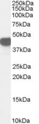
Supportive validation
- Submitted by
- Novus Biologicals (provider)
- Main image

- Experimental details
- Western Blot: Argininosuccinate Synthase Antibody [NBP1-00153] - Analysis of Argininosuccinate Synthase in Human Kidney lysate (35ug protein in RIPA buffer) using this antibody at 0.03ug/ml. Primary incubation was 1 hour. Detected by chemiluminescence.
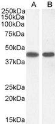
- Submitted by
- Novus Biologicals (provider)
- Main image

- Experimental details
- Western Blot: Argininosuccinate Synthase Antibody [NBP1-00153] - Staining of A431 (A) and (1ug/ml) NIH3T3(B) cell lysate (35 ug protein in RIPA buffer). Antibody at 0.3 ug/mL. Detected by chemiluminescence.
- Submitted by
- Novus Biologicals (provider)
- Main image
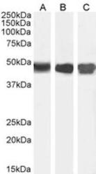
- Experimental details
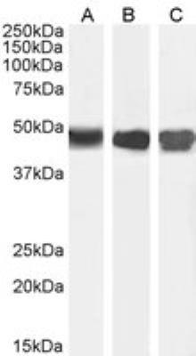
- Western Blot: Argininosuccinate Synthase Antibody [NBP1-00153] - Staining of Human Kidney (A) Mouse Liver (B) with antibody at 0.01 ug/mL and Rat Kidney (C) lysate with antibody at 0.03 ug/mL (35 ug protein in RIPA buffer). Detected by chemiluminescence.
Supportive validation
- Submitted by
- Novus Biologicals (provider)
- Main image
- Experimental details
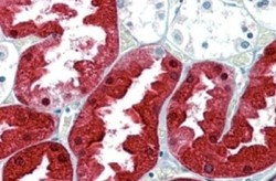
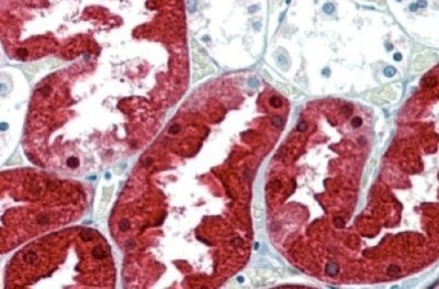
- Immunohistochemistry-Paraffin: Argininosuccinate Synthase Antibody [NBP1-00153] - (2.5ug/ml) staining of paraffin embedded Human Kidney. Steamed antigen retrieval with citrate buffer pH 6, AP-staining.
- Submitted by
- Novus Biologicals (provider)
- Main image
- Experimental details
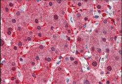
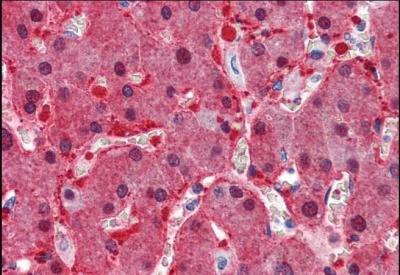
- Immunohistochemistry-Paraffin: Argininosuccinate Synthase Antibody [NBP1-00153] - (2.5ug/ml) staining of paraffin embedded Human Liver. Steamed antigen retrieval with citrate buffer pH 6, AP-staining.
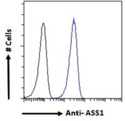
Supportive validation
- Submitted by
- Novus Biologicals (provider)
- Main image

- Experimental details
- Flow Cytometry: Argininosuccinate Synthase Antibody [NBP1-00153] - Analysis of paraformaldehyde fixed A431 cells (blue line), permeabilized with 0.5% Triton. Primary incubation 1hr (10 ug/mL) followed by Alexa Fluor 488 secondary antibody (1 ug/mL). IgG control: Unimmunized goat IgG (black line) followed by Alexa Fluor 488 secondary antibody.