 Explore
 Validate
 Learn
Learn Western blot
 Immunocytochemistry
Antibody data
- Antibody Data
- Antigen structure
- References [1]
- Comments [0]
- Validations
- Immunocytochemistry [3]
- Immunohistochemistry [3]
Submit
Validation data
Reference
Comment
Report error
- Product number
- GTX22871 - Provider product page

- Provider
- GeneTex
- Proper citation
- GeneTex Cat#GTX22871, RRID:AB_384890
- Product name
- Sodium/Potassium ATPase alpha 1 antibody [M7-PB-E9]
- Antibody type
- Monoclonal
- Reactivity
- Human, Mouse, Rat, Bovine, Canine, Chicken/Avian, Porcine, Sheep
- Host
- Mouse
Submitted references A zebrafish chemical suppressor screening identifies small molecule inhibitors of the Wnt/β-catenin pathway.
Nishiya N, Oku Y, Kumagai Y, Sato Y, Yamaguchi E, Sasaki A, Shoji M, Ohnishi Y, Okamoto H, Uehara Y
Chemistry & biology 2014 Apr 24;21(4):530-540
Chemistry & biology 2014 Apr 24;21(4):530-540
No comments: Submit comment
Supportive validation
- Submitted by
- GeneTex (provider)
- Main image
- Experimental details
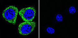
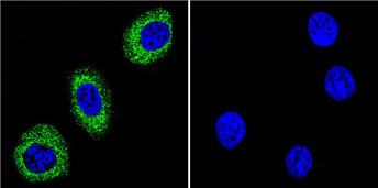
- Immunofluorescent analysis of Sodium/Potassium ATPase alpha using Sodium/Potassium ATPase alpha Monoclonal Antibody (M7-PB-E9) (GTX22871) shows staining in Hela Cells. Sodium/Potassium ATPase alpha (green), F-Actin staining with Phalloidin (red) and nuclei with DAPI (blue) is shown. Cells were grown on chamber slides and fixed with formaldehyde prior to staining. Cells were probed without (control) or with an antibody recognizing Sodium/Potassium ATPase alpha (GTX22871) at a dilution of 1:20 over night at 4 degrees C, washed with PBS and incubated with a DyLight-488 conjugated secondary antibody. Images were taken at 60X magnification.

- Submitted by
- GeneTex (provider)
- Main image
- Experimental details
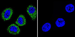
- Immunofluorescent analysis of Sodium/Potassium ATPase alpha using Sodium/Potassium ATPase alpha Monoclonal Antibody (M7-PB-E9) (GTX22871) shows staining in MCF-7 Cells. Sodium/Potassium ATPase alpha (green), F-Actin staining with Phalloidin (red) and nuclei with DAPI (blue) is shown. Cells were grown on chamber slides and fixed with formaldehyde prior to staining. Cells were probed without (control) or with an antibody recognizing Sodium/Potassium ATPase alpha (GTX22871) at a dilution of 1:20 over night at 4 degrees C, washed with PBS and incubated with a DyLight-488 conjugated secondary antibody. Images were taken at 60X magnification.
- Submitted by
- GeneTex (provider)
- Main image
- Experimental details
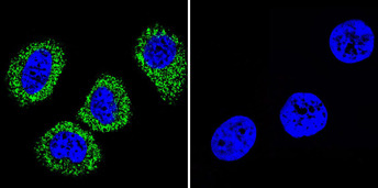
- Immunofluorescent analysis of Sodium/Potassium ATPase alpha using Sodium/Potassium ATPase alpha Monoclonal Antibody (M7-PB-E9) (GTX22871) shows staining in U251 Cells. Sodium/Potassium ATPase alpha (green), F-Actin staining with Phalloidin (red) and nuclei with DAPI (blue) is shown. Cells were grown on chamber slides and fixed with formaldehyde prior to staining. Cells were probed without (control) or with an antibody recognizing Sodium/Potassium ATPase alpha (GTX22871) at a dilution of 1:20 over night at 4 degrees C, washed with PBS and incubated with a DyLight-488 conjugated secondary antibody. Images were taken at 60X magnification.
Supportive validation
- Submitted by
- GeneTex (provider)
- Main image

- Experimental details
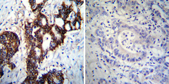
- Immunohistochemistry was performed on cancer biopsies of deparaffinized Human colon carcinoma tissues. To expose target proteins, heat induced antigen retrieval was performed using 10mM sodium citrate (pH6.0) buffer, microwaved for 8-15 minutes. Following antigen retrieval tissues were blocked in 3% BSA-PBS for 30 minutes at room temperature. Tissues were then probed at a dilution of 1:100 with a mouse monoclonal antibody recognizing Sodium/Potassium ATPase alpha (GTX22871) or without primary antibody (negative control) overnight at 4¢XC in a humidified chamber. Tissues were washed extensively with PBST and endogenous peroxidase activity was quenched with a peroxidase suppressor. Detection was performed using a biotin-conjugated secondary antibody and SA-HRP, followed by colorimetric detection using DAB. Tissues were counterstained with hematoxylin and prepped for mounting.
- Submitted by
- GeneTex (provider)
- Main image
- Experimental details
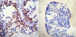
- Immunohistochemistry was performed on normal deparaffinized Human testis tissue tissues. To expose target proteins, heat induced antigen retrieval was performed using 10mM sodium citrate (pH6.0) buffer, microwaved for 8-15 minutes. Following antigen retrieval tissues were blocked in 3% BSA-PBS for 30 minutes at room temperature. Tissues were then probed at a dilution of 1:200 with a mouse monoclonal antibody recognizing Sodium/Potassium ATPase alpha (GTX22871) or without primary antibody (negative control) overnight at 4¢XC in a humidified chamber. Tissues were washed extensively with PBST and endogenous peroxidase activity was quenched with a peroxidase suppressor. Detection was performed using a biotin-conjugated secondary antibody and SA-HRP, followed by colorimetric detection using DAB. Tissues were counterstained with hematoxylin and prepped for mounting.
- Submitted by
- GeneTex (provider)
- Main image
- Experimental details
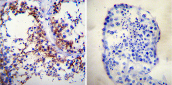
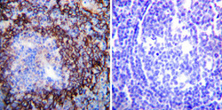
- Immunohistochemistry was performed on normal deparaffinized Human tonsil tissue tissues. To expose target proteins, heat induced antigen retrieval was performed using 10mM sodium citrate (pH6.0) buffer, microwaved for 8-15 minutes. Following antigen retrieval tissues were blocked in 3% BSA-PBS for 30 minutes at room temperature. Tissues were then probed at a dilution of 1:200 with a mouse monoclonal antibody recognizing Sodium/Potassium ATPase alpha (GTX22871) or without primary antibody (negative control) overnight at 4¢XC in a humidified chamber. Tissues were washed extensively with PBST and endogenous peroxidase activity was quenched with a peroxidase suppressor. Detection was performed using a biotin-conjugated secondary antibody and SA-HRP, followed by colorimetric detection using DAB. Tissues were counterstained with hematoxylin and prepped for mounting.
