Explore
 Validate
 Learn
LearnPA1-213A
antibody from Invitrogen Antibodies
Targeting: THRB
ERBA-BETA, ERBA2, GRTH, NR1A2, PRTH, THR1, THRB1, THRB2
 Western blot
 Immunocytochemistry
Antibody data
- Antibody Data
- Antigen structure
- References [12]
- Comments [0]
- Validations
- Immunocytochemistry [5]
- Immunohistochemistry [3]
- Other assay [2]
Submit
Validation data
Reference
Comment
Report error
- Product number
- PA1-213A - Provider product page

- Provider
- Invitrogen Antibodies
- Product name
- THRB Polyclonal Antibody
- Antibody type
- Polyclonal
- Antigen
- Synthetic peptide
- Description
- PA1-213A detects thyroid hormone receptor (TR) beta-1 from human and rat tissues. This antibody does not detect TR alpha-1 or TRv alpha-2. PA1-213A has been successfully used in Western blot, immunohistochemistry (paraffin), IF and gel shift procedures. By Western blot, this antibody detects a ~55 kDa protein representing recombinant human TRv beta-1 expressed in E. coli. PA1-213A immunizing peptide corresponds to residues 62-81 of human TR beta-1, with an N-terminal added cysteine. Alternative numbering of this protein has this peptide corresponding to residues 67-86 of human TR beta-1. This sequence is completely conserved between human and rat TR beta-1.
- Reactivity
- Human, Rat
- Host
- Rabbit
- Isotype
- IgG
- Vial size
- 100 μL
- Concentration
- Conc. Not Determined
- Storage
- -20°C, Avoid Freeze/Thaw Cycles
Submitted references Thyroid hormone inhibits lung fibrosis in mice by improving epithelial mitochondrial function.
Thyroid hormone receptor regulates most genes independently of fibroblast growth factor 21 in liver.
Impaired hair growth and wound healing in mice lacking thyroid hormone receptors.
Barhl1 is directly regulated by thyroid hormone in the developing cerebellum of mice.
Thyroid hormone-regulated gene expression in juvenile mouse liver: identification of thyroid response elements using microarray profiling and in silico analyses.
Aging impairs myocardial fatty acid and ketone oxidation and modifies cardiac functional and metabolic responses to insulin in mice.
The dominant negative thyroid hormone receptor beta-mutant {Delta}337T alters PPAR{alpha} signaling in heart.
Multiple messenger ribonucleic acid variants regulate cell-specific expression of human thyroid hormone receptor beta1.
TR expression and function in human bone marrow stromal and osteoblast-like cells.
Expression of thyroid receptor isoforms in the human fetal central nervous system and the effects of intrauterine growth restriction.
Expression of thyroid receptor isoforms in the human fetal central nervous system and the effects of intrauterine growth restriction.
Antipeptide polyclonal antibodies specifically recognize each human thyroid hormone receptor isoform.
Yu G, Tzouvelekis A, Wang R, Herazo-Maya JD, Ibarra GH, Srivastava A, de Castro JPW, DeIuliis G, Ahangari F, Woolard T, Aurelien N, Arrojo E Drigo R, Gan Y, Graham M, Liu X, Homer RJ, Scanlan TS, Mannam P, Lee PJ, Herzog EL, Bianco AC, Kaminski N
Nature medicine 2018 Jan;24(1):39-49
Nature medicine 2018 Jan;24(1):39-49
Thyroid hormone receptor regulates most genes independently of fibroblast growth factor 21 in liver.
Zhang A, Sieglaff DH, York JP, Suh JH, Ayers SD, Winnier GE, Kharitonenkov A, Pin C, Zhang P, Webb P, Xia X
The Journal of endocrinology 2015 Mar;224(3):289-301
The Journal of endocrinology 2015 Mar;224(3):289-301
Impaired hair growth and wound healing in mice lacking thyroid hormone receptors.
Contreras-Jurado C, García-Serrano L, Martínez-Fernández M, Ruiz-Llorente L, Paramio JM, Aranda A
PloS one 2014;9(9):e108137
PloS one 2014;9(9):e108137
Barhl1 is directly regulated by thyroid hormone in the developing cerebellum of mice.
Dong H, Yauk CL, Wade MG
Biochemical and biophysical research communications 2011 Nov 11;415(1):157-62
Biochemical and biophysical research communications 2011 Nov 11;415(1):157-62
Thyroid hormone-regulated gene expression in juvenile mouse liver: identification of thyroid response elements using microarray profiling and in silico analyses.
Paquette MA, Dong H, Gagné R, Williams A, Malowany M, Wade MG, Yauk CL
BMC genomics 2011 Dec 29;12:634
BMC genomics 2011 Dec 29;12:634
Aging impairs myocardial fatty acid and ketone oxidation and modifies cardiac functional and metabolic responses to insulin in mice.
Hyyti OM, Ledee D, Ning XH, Ge M, Portman MA
American journal of physiology. Heart and circulatory physiology 2010 Sep;299(3):H868-75
American journal of physiology. Heart and circulatory physiology 2010 Sep;299(3):H868-75
The dominant negative thyroid hormone receptor beta-mutant {Delta}337T alters PPAR{alpha} signaling in heart.
Buroker NE, Young ME, Wei C, Serikawa K, Ge M, Ning XH, Portman MA
American journal of physiology. Endocrinology and metabolism 2007 Feb;292(2):E453-60
American journal of physiology. Endocrinology and metabolism 2007 Feb;292(2):E453-60
Multiple messenger ribonucleic acid variants regulate cell-specific expression of human thyroid hormone receptor beta1.
Frankton S, Harvey CB, Gleason LM, Fadel A, Williams GR
Molecular endocrinology (Baltimore, Md.) 2004 Jul;18(7):1631-42
Molecular endocrinology (Baltimore, Md.) 2004 Jul;18(7):1631-42
TR expression and function in human bone marrow stromal and osteoblast-like cells.
Siddiqi A, Parsons MP, Lewis JL, Monson JP, Williams GR, Burrin JM
The Journal of clinical endocrinology and metabolism 2002 Feb;87(2):906-14
The Journal of clinical endocrinology and metabolism 2002 Feb;87(2):906-14
Expression of thyroid receptor isoforms in the human fetal central nervous system and the effects of intrauterine growth restriction.
Kilby MD, Gittoes N, McCabe C, Verhaeg J, Franklyn JA
Clinical endocrinology 2000 Oct;53(4):469-77
Clinical endocrinology 2000 Oct;53(4):469-77
Expression of thyroid receptor isoforms in the human fetal central nervous system and the effects of intrauterine growth restriction.
Kilby MD, Gittoes N, McCabe C, Verhaeg J, Franklyn JA
Clinical endocrinology 2000 Oct;53(4):469-77
Clinical endocrinology 2000 Oct;53(4):469-77
Antipeptide polyclonal antibodies specifically recognize each human thyroid hormone receptor isoform.
Falcone M, Miyamoto T, Fierro-Renoy F, Macchia E, DeGroot LJ
Endocrinology 1992 Nov;131(5):2419-29
Endocrinology 1992 Nov;131(5):2419-29
No comments: Submit comment
Supportive validation
- Submitted by
- Invitrogen Antibodies (provider)
- Main image
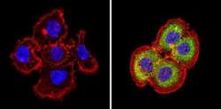
- Experimental details
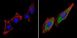
- Immunofluorescent analysis of Thyroid Hormone Receptor beta-1 (green) showing staining in the cytoplasm of A431 cells (right) compared to a negative control without primary antibody (left). Formalin-fixed cells were permeabilized with 0.1% Triton X-100 in TBS for 5-10 minutes and blocked with 3% BSA-PBS for 30 minutes at room temperature. Cells were probed with a Thyroid Hormone Receptor beta-1 polyclonal antibody (Product # PA1-213A) in 3% BSA-PBS at a dilution of 1:100 and incubated overnight at 4ºC in a humidified chamber. Cells were washed with PBST and incubated with a DyLight-conjugated secondary antibody in PBS at room temperature in the dark. F-actin (red) was stained with a fluorescent red phalloidin and nuclei (blue) were stained with Hoechst or DAPI. Images were taken at a magnification of 60x.
- Submitted by
- Invitrogen Antibodies (provider)
- Main image

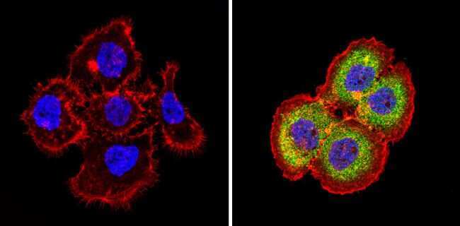
- Experimental details
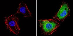
- Immunofluorescent analysis of Thyroid Hormone Receptor beta-1 (green) showing staining in the cytoplasm and nucleus of Hela cells (right) compared to a negative control without primary antibody (left). Formalin-fixed cells were permeabilized with 0.1% Triton X-100 in TBS for 5-10 minutes and blocked with 3% BSA-PBS for 30 minutes at room temperature. Cells were probed with a Thyroid Hormone Receptor beta-1 polyclonal antibody (Product # PA1-213A) in 3% BSA-PBS at a dilution of 1:100 and incubated overnight at 4ºC in a humidified chamber. Cells were washed with PBST and incubated with a DyLight-conjugated secondary antibody in PBS at room temperature in the dark. F-actin (red) was stained with a fluorescent red phalloidin and nuclei (blue) were stained with Hoechst or DAPI. Images were taken at a magnification of 60x.
- Submitted by
- Invitrogen Antibodies (provider)
- Main image
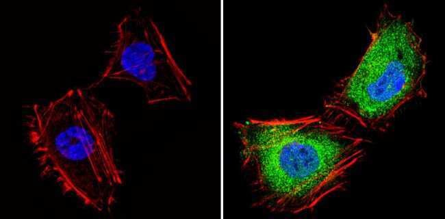
- Experimental details
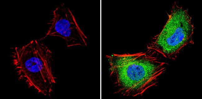
- Immunofluorescent analysis of Thyroid Hormone Receptor beta-1 (green) showing staining in the cytoplasm of L6 cells (right) compared to a negative control without primary antibody (left). Formalin-fixed cells were permeabilized with 0.1% Triton X-100 in TBS for 5-10 minutes and blocked with 3% BSA-PBS for 30 minutes at room temperature. Cells were probed with a Thyroid Hormone Receptor beta-1 polyclonal antibody (Product # PA1-213A) in 3% BSA-PBS at a dilution of 1:100 and incubated overnight at 4ºC in a humidified chamber. Cells were washed with PBST and incubated with a DyLight-conjugated secondary antibody in PBS at room temperature in the dark. F-actin (red) was stained with a fluorescent red phalloidin and nuclei (blue) were stained with Hoechst or DAPI. Images were taken at a magnification of 60x.

- Submitted by
- Invitrogen Antibodies (provider)
- Main image
- Experimental details
- Immunofluorescent analysis of Thyroid Hormone Receptor beta-1 (green) showing staining in the cytoplasm and nucleus of Hela cells (right) compared to a negative control without primary antibody (left). Formalin-fixed cells were permeabilized with 0.1% Triton X-100 in TBS for 5-10 minutes and blocked with 3% BSA-PBS for 30 minutes at room temperature. Cells were probed with a Thyroid Hormone Receptor beta-1 polyclonal antibody (Product # PA1-213A) in 3% BSA-PBS at a dilution of 1:100 and incubated overnight at 4ºC in a humidified chamber. Cells were washed with PBST and incubated with a DyLight-conjugated secondary antibody in PBS at room temperature in the dark. F-actin (red) was stained with a fluorescent red phalloidin and nuclei (blue) were stained with Hoechst or DAPI. Images were taken at a magnification of 60x.
- Submitted by
- Invitrogen Antibodies (provider)
- Main image

- Experimental details
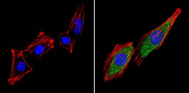
- Immunofluorescent analysis of Thyroid Hormone Receptor beta-1 (green) showing staining in the cytoplasm of L6 cells (right) compared to a negative control without primary antibody (left). Formalin-fixed cells were permeabilized with 0.1% Triton X-100 in TBS for 5-10 minutes and blocked with 3% BSA-PBS for 30 minutes at room temperature. Cells were probed with a Thyroid Hormone Receptor beta-1 polyclonal antibody (Product # PA1-213A) in 3% BSA-PBS at a dilution of 1:100 and incubated overnight at 4ºC in a humidified chamber. Cells were washed with PBST and incubated with a DyLight-conjugated secondary antibody in PBS at room temperature in the dark. F-actin (red) was stained with a fluorescent red phalloidin and nuclei (blue) were stained with Hoechst or DAPI. Images were taken at a magnification of 60x.
Supportive validation
- Submitted by
- Invitrogen Antibodies (provider)
- Main image
- Experimental details
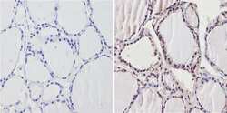
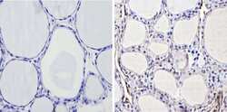
- Immunohistochemistry analysis of Thyroid Hormone Receptor beta-1 showing positive staining in the nucleus of paraffin-treated Human thyroid tissue (right) compared with a negative control in the absence of primary antibody (left). To expose target proteins, antigen retrieval method was performed using 10mM sodium citrate (pH 6.0) microwaved for 8-15 min. Following antigen retrieval, tissues were blocked in 3% H2O2-methanol for 15 min at room temperature, washed with ddH2O and PBS, and then probed with a Thyroid Hormone Receptor beta-1 polyclonal antibody (Product # PA1-213A) diluted by 3% BSA-PBS at a dilution of 1:200 overnight at 4°C in a humidified chamber. Tissues were washed extensively PBST and detection was performed using an HRP-conjugated secondary antibody followed by colorimetric detection using a DAB kit. Tissues were counterstained with hematoxylin and dehydrated with ethanol and xylene to prep for mounting.
- Submitted by
- Invitrogen Antibodies (provider)
- Main image
- Experimental details
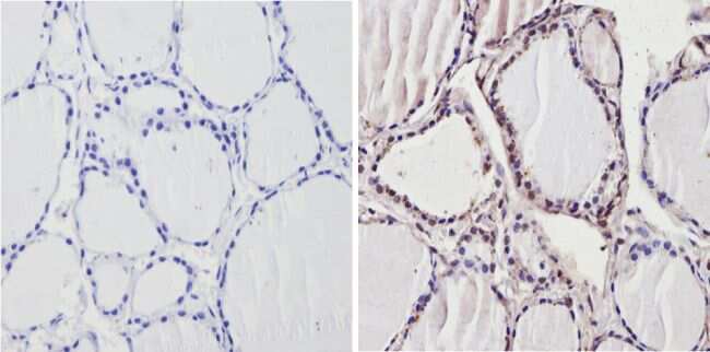
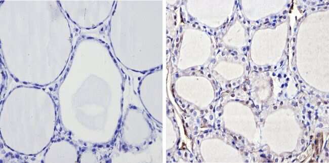
- Immunohistochemistry analysis of Thyroid Hormone Receptor beta-1 showing positive staining in the nucleus of paraffin-treated Rat thyroid tissue (right) compared with a negative control in the absence of primary antibody (left). To expose target proteins, antigen retrieval method was performed using 10mM sodium citrate (pH 6.0) microwaved for 8-15 min. Following antigen retrieval, tissues were blocked in 3% H2O2-methanol for 15 min at room temperature, washed with ddH2O and PBS, and then probed with a Thyroid Hormone Receptor beta-1 polyclonal antibody (Product # PA1-213A) diluted by 3% BSA-PBS at a dilution of 1:200 overnight at 4°C in a humidified chamber. Tissues were washed extensively PBST and detection was performed using an HRP-conjugated secondary antibody followed by colorimetric detection using a DAB kit. Tissues were counterstained with hematoxylin and dehydrated with ethanol and xylene to prep for mounting.
- Submitted by
- Invitrogen Antibodies (provider)
- Main image
- Experimental details
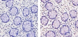
- Immunohistochemistry analysis of Thyroid Hormone Receptor beta-1 showing staining in the nucleus and weak staining in the cytoplasm of paraffin-embedded human colon tissue (right) compared to a negative control without primary antibody (left). To expose target proteins, antigen retrieval was performed using 10mM sodium citrate (pH 6.0), microwaved for 8-15 min. Following antigen retrieval, tissues were blocked in 3% H2O2-methanol for 15 min at room temperature, washed with ddH2O and PBS, and then probed with a Thyroid Hormone Receptor beta-1 Rabbit Polyclonal Antibody (Product # PA1-213A) diluted in 3% BSA-PBS at a dilution of 1:20 for 1 hour at 37ºC in a humidified chamber. Tissues were washed extensively in PBST and detection was performed using an HRP-conjugated secondary antibody followed by colorimetric detection using a DAB kit. Tissues were counterstained with hematoxylin and dehydrated with ethanol and xylene to prep for mounting.

Supportive validation
- Submitted by
- Invitrogen Antibodies (provider)
- Main image
- Experimental details
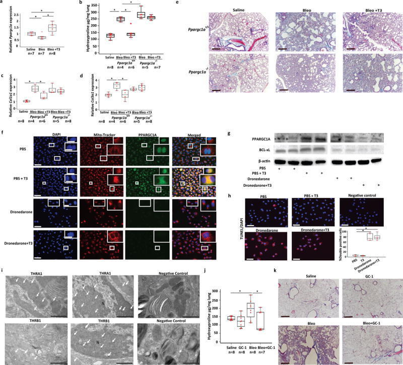
- Figure 5 Anti-fibrotic effects of TH are mediated through PPARGC1A. ( a )Quantitative RT-PCR analysis for Ppargc1a mRNA levels in theindicated treatment groups (means + SEM),* P < 0.001. ( b ) Lunghydroxyproline content, and quantitative RT-PCR analysis of collagen type 1,alpha 1 ( Col1a1 ) (c) and type 3, alpha 1( Col3a1 ) (d) mRNA levels in Ppargc1a -deficient( Ppargc1a -/- ) mice or wild-typelittermates ( Ppargc1a +/+ ) treated with aerosolized T3 followingintratracheal challenge with bleomycin or equivalent volume of normal saline.Data presented are from one of two independent experiments with similar resultsand are expressed as mean hydroxyproline content per lung (mug/gr lung)set + SEM, * P
- Submitted by
- Invitrogen Antibodies (provider)
- Main image
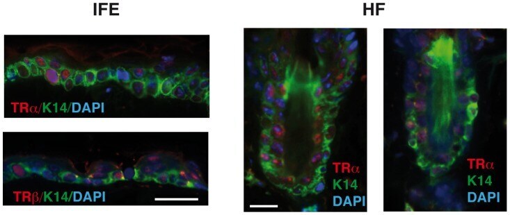
- Experimental details
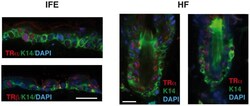
- Figure 3 Both TRalpha and TRbeta are expressed in the mouse skin. Double immunofluorescence images of expression of keratin 14 (K14, green) with TRalpha or TRbeta (red). Images were obtained from the interfollicular epidermis (IFE) and the hair follicles (HF) of wild-type mice. The slides were counterstained with DAPI (blue) and the merged images are shown. Bars: 50 uM.