Explore
 Validate
 Learn
Learn Western blot
Antibody data
- Antibody Data
- Antigen structure
- References [3]
- Comments [0]
- Validations
- Western blot [1]
- Immunohistochemistry [3]
- Other assay [1]
Submit
Validation data
Reference
Comment
Report error
- Product number
- AHO1362 - Provider product page

- Provider
- Invitrogen Antibodies
- Product name
- JNK1/JNK2 Monoclonal Antibody (279Q38)
- Antibody type
- Monoclonal
- Antigen
- Recombinant protein fragment
- Description
- Recommended positive controls: human Jurkat cells, mouse L929 cells and rat L6 cells.
- Antibody clone number
- 279Q38
- Concentration
- 0.5 mg/mL
Submitted references Progesterone suppresses the progression of colonic carcinoma by increasing the activity of the GADD45α/JNK/c‑Jun signalling pathway.
Molecular mechanism of mice gastric oxidative damage induced by nanoparticulate titanium dioxide.
Nanoparticulate titanium dioxide-inhibited dendritic development is involved in apoptosis and autophagy of hippocampal neurons in offspring mice.
Zhang YL, Wen XD, Guo X, Huang SQ, Wang TT, Zhou PT, Li W, Zhou LF, Hu YH
Oncology reports 2021 Jun;45(6)
Oncology reports 2021 Jun;45(6)
Molecular mechanism of mice gastric oxidative damage induced by nanoparticulate titanium dioxide.
Ji J, Zhou Y, Hong F, Ze Y, Fan D, Zhang X
Toxicology research 2021 Jan;10(1):60-67
Toxicology research 2021 Jan;10(1):60-67
Nanoparticulate titanium dioxide-inhibited dendritic development is involved in apoptosis and autophagy of hippocampal neurons in offspring mice.
Zhou Y, Hong F, Tian Y, Zhao X, Hong J, Ze Y, Wang L
Toxicology research 2017 Nov 1;6(6):889-901
Toxicology research 2017 Nov 1;6(6):889-901
No comments: Submit comment
Supportive validation
- Submitted by
- Invitrogen Antibodies (provider)
- Main image
- Experimental details
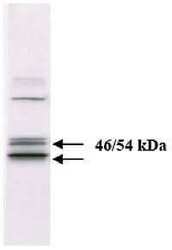
- Proteins from cell extract of human Jurkat cells were resolved by SDS-PAGE and transferred to PVDF. The membranes were incubated with this JNK1/2 monoclonal antibody (clone 279Q38) at a concentration of 1 µg/mL for two hours at room temperature

Supportive validation
- Submitted by
- Invitrogen Antibodies (provider)
- Main image
- Experimental details
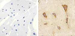
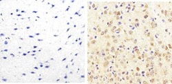
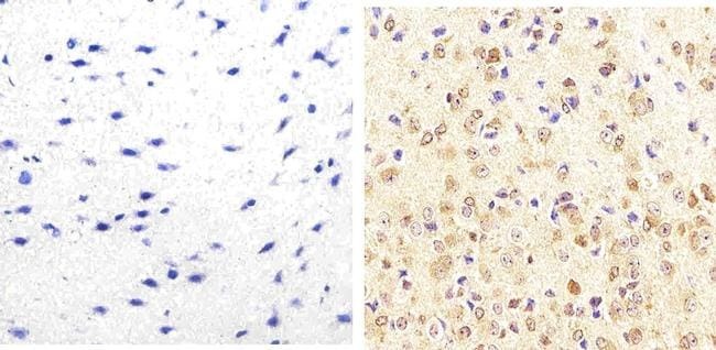
- Immunohistochemistry analysis of JNK1/2 showing staining in the cytoplasm and nucleus of paraffin-embedded human brain tissue (right) compared to a negative control without primary antibody (left). To expose target proteins, antigen retrieval was performed using 10mM sodium citrate (pH 6.0), microwaved for 8-15 min. Following antigen retrieval, tissues were blocked in 3% H2O2-methanol for 15 min at room temperature, washed with ddH2O and PBS, and then probed with a JNK1/2 monoclonal antibody (Product # AHO1362) diluted in 3% BSA-PBS at a dilution of 1:20 overnight at 4ºC in a humidified chamber. Tissues were washed extensively in PBST and detection was performed using an HRP-conjugated secondary antibody followed by colorimetric detection using a DAB kit. Tissues were counterstained with hematoxylin and dehydrated with ethanol and xylene to prep for mounting.
- Submitted by
- Invitrogen Antibodies (provider)
- Main image
- Experimental details
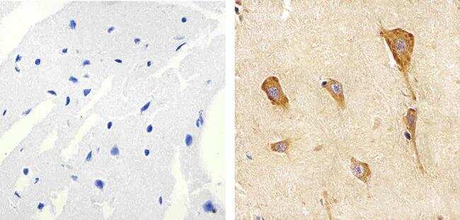
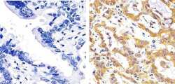
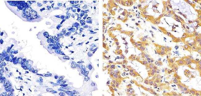
- Immunohistochemistry analysis of JNK1/2 showing staining in the cytoplasm and nucleus of paraffin-embedded human lung adenocarcinoma tissue (right) compared to a negative control without primary antibody (left). To expose target proteins, antigen retrieval was performed using 10mM sodium citrate (pH 6.0), microwaved for 8-15 min. Following antigen retrieval, tissues were blocked in 3% H2O2-methanol for 15 min at room temperature, washed with ddH2O and PBS, and then probed with a JNK1/2 monoclonal antibody (Product # AHO1362) diluted in 3% BSA-PBS at a dilution of 1:20 overnight at 4ºC in a humidified chamber. Tissues were washed extensively in PBST and detection was performed using an HRP-conjugated secondary antibody followed by colorimetric detection using a DAB kit. Tissues were counterstained with hematoxylin and dehydrated with ethanol and xylene to prep for mounting.
- Submitted by
- Invitrogen Antibodies (provider)
- Main image
- Experimental details
- Immunohistochemistry analysis of JNK1/2 showing staining in the cytoplasm and nucleus of paraffin-embedded mouse brain tissue (right) compared to a negative control without primary antibody (left). To expose target proteins, antigen retrieval was performed using 10mM sodium citrate (pH 6.0), microwaved for 8-15 min. Following antigen retrieval, tissues were blocked in 3% H2O2-methanol for 15 min at room temperature, washed with ddH2O and PBS, and then probed with a JNK1/2 monoclonal antibody (Product # AHO1362) diluted in 3% BSA-PBS at a dilution of 1:20 overnight at 4ºC in a humidified chamber. Tissues were washed extensively in PBST and detection was performed using an HRP-conjugated secondary antibody followed by colorimetric detection using a DAB kit. Tissues were counterstained with hematoxylin and dehydrated with ethanol and xylene to prep for mounting.
Supportive validation
- Submitted by
- Invitrogen Antibodies (provider)
- Main image
- Experimental details
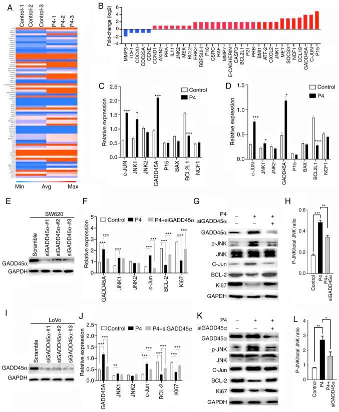
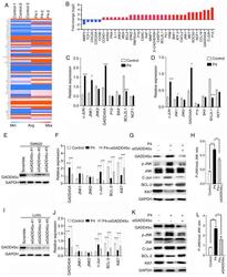
- Figure 5. Progesterone up-regulates the JNK pathway via GADD45alpha activation to inhibit progression of colonic carcinoma. (A) Heat map depicting progesterone-induced changes in the expression profile of genes that were assessed on the PCR microarray. Blue and red represent low and high gene expression levels in pinnae, respectively. (B) Selection of genes that were altered the most in SW620 cells. Genes are presented alphabetically. RT-PCR was used to investigate the effect of progesterone on proliferation-related genes such as c-Jun, JNK1, JNK2, GADD45alpha, P15, BAX, BCL2L1 and NCF1 in (C) SW620 and (D) LoVo cells Efficiency of knockdown GADD45alpha in (E) SW620 and (I) LoVo cells, respectively. Expression of GADD45alpha, JNK1, JNK2, c-Jun, BCL-2, and Ki67 in (F) SW620 and (J) LoVo cells was analysed using RT-PCR, and GADD45alpha, phosphorylation of JNK, JNK, c-Jun, BCL-2 and Ki67 in (G) SW620 and (K) LoVo cells were analysed using western blot. Relative expression of phosphorylation of JNK to total protein of JNK in (H) SW620 and (L) LoVo cells. *P