Explore
 Validate
 Learn
Learn Western blot
Antibody data
- Antibody Data
- Antigen structure
- References [2]
- Comments [0]
- Validations
- Western blot [2]
- Immunocytochemistry [1]
Submit
Validation data
Reference
Comment
Report error
- Product number
- MAB1846 - Provider product page

- Provider
- R&D Systems
- Product name
- Human/Mouse/Rat JNK2 Antibody
- Antibody type
- Monoclonal
- Description
- Protein A or G purified from hybridoma culture supernatant. Detects human, mouse, and rat JNK2, expressed as p46 JNK (isoforms 2 and/or 3, both 382 aa) and p54 JNK (isoforms 1 and/or 4, both 424 aa) in Western blots. Does not detect recombinant JNK1 or JNK3.
- Reactivity
- Human, Mouse, Rat
- Host
- Mouse
- Conjugate
- Unconjugated
- Isotype
- IgG
- Antibody clone number
- 252320
- Vial size
- 100 ug
- Concentration
- LYOPH
- Storage
- Use a manual defrost freezer and avoid repeated freeze-thaw cycles. 12 months from date of receipt, -20 to -70 °C as supplied. 1 month, 2 to 8 °C under sterile conditions after reconstitution. 6 months, -20 to -70 °C under sterile conditions after reconstitution.
Submitted references APP upregulation contributes to retinal ganglion cell degeneration via JNK3.
Distinct role of c-Jun N-terminal kinase isoforms in human neutrophil apoptosis regulated by tumor necrosis factor-alpha and granulocyte-macrophage colony-stimulating factor.
Liu C, Zhang CW, Zhou Y, Wong WQ, Lee LC, Ong WY, Yoon SO, Hong W, Fu XY, Soong TW, Koo EH, Stanton LW, Lim KL, Xiao ZC, Dawe GS
Cell death and differentiation 2018 Mar;25(4):663-678
Cell death and differentiation 2018 Mar;25(4):663-678
Distinct role of c-Jun N-terminal kinase isoforms in human neutrophil apoptosis regulated by tumor necrosis factor-alpha and granulocyte-macrophage colony-stimulating factor.
Kato T, Noma H, Kitagawa M, Takahashi T, Oshitani N, Kitagawa S
Journal of interferon & cytokine research : the official journal of the International Society for Interferon and Cytokine Research 2008 Apr;28(4):235-43
Journal of interferon & cytokine research : the official journal of the International Society for Interferon and Cytokine Research 2008 Apr;28(4):235-43
No comments: Submit comment
Supportive validation
- Submitted by
- R&D Systems (provider)
- Main image

- Experimental details
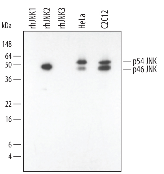
- Detection of Human, Mouse, and Rat JNK2 by Western Blot. Western blot shows lysates of HeLa human cervical epithelial carcinoma cell line and C2C12 mouse myoblast cell line. PVDF membrane was probed with 0.2 µg/mL Mouse Anti-Human/Mouse/Rat JNK2 Monoclonal Antibody (Catalog # MAB1846) followed by HRP-conjugated Anti-Mouse IgG Secondary Antibody (Catalog # HAF007). For additional reference, recombinant human JNK1, JNK2, and JNK3 (1 ng/lane) were included. Specific bands for JNK2 were detected at approximately 46 and 54 kDa (as indicated). This experiment was conducted under reducing conditions and using Immunoblot Buffer Group 1.
- Submitted by
- R&D Systems (provider)
- Main image
- Experimental details
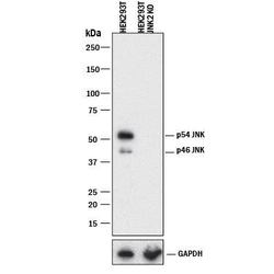
- Western Blot Shows Human JNK2 Specificity by Using Knockout Cell Line. Western blot shows lysates of HEK293T human embryonic kidney parental cell line and JNK2 knockout HEK293T cell line (KO). PVDF membrane was probed with 0.2 µg/mL of Mouse Anti-Human/Mouse/Rat JNK2 Monoclonal Antibody (Catalog # MAB1846) followed by HRP-conjugated Anti-Mouse IgG Secondary Antibody (Catalog # HAF018). Specific bands were detected for JNK2 at approximately 45 and 54 kDa (as indicated) in the parental HEK293T cell line, but is not detectable in knockout HEK293T cell line. GAPDH (Catalog # MAB5718) is shown as a loading control. This experiment was conducted under reducing conditions and using Immunoblot Buffer Group 1.
Supportive validation
- Submitted by
- R&D Systems (provider)
- Main image
- Experimental details
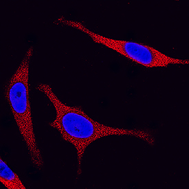
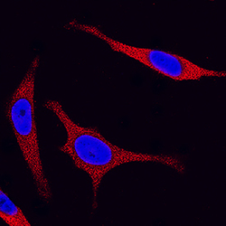
- JNK2 in HeLa Human Cell Line. JNK2 was detected in immersion fixed HeLa human cervical epithelial carcinoma cell line using Mouse Anti-Human/Mouse/Rat JNK2 Monoclonal Antibody (Catalog # MAB1846) at 25 µg/mL for 3 hours at room temperature. Cells were stained using the NorthernLights™ 557-conjugated Anti-Mouse IgG Secondary Antibody (red; Catalog # NL007) and counterstained with DAPI (blue). Specific staining was localized to cytoplasm. View our protocol for Fluorescent ICC Staining of Cells on Coverslips.