Explore
 Validate
 Learn
Learn Western blot
 Immunocytochemistry
Antibody data
- Antibody Data
- Antigen structure
- References [1]
- Comments [0]
- Validations
- Western blot [7]
- Immunohistochemistry [4]
Submit
Validation data
Reference
Comment
Report error
- Product number
- NBP2-19459 - Provider product page

- Provider
- Novus Biologicals
- Product name
- Rabbit Polyclonal N-Cadherin Antibody
- Antibody type
- Polyclonal
- Description
- Immunogen affinity purified.
- Reactivity
- Human, Mouse, Rat
- Host
- Rabbit
- Isotype
- IgG
- Vial size
- 0.1 ml
- Storage
- Aliquot and store at -20C or -80C. Avoid freeze-thaw cycles.
Submitted references Improved ovarian cancer EMT-CTC isolation by immunomagnetic targeting of epithelial EpCAM and mesenchymal N-cadherin.
Po JW, Roohullah A, Lynch D, DeFazio A, Harrison M, Harnett PR, Kennedy C, de Souza P, Becker TM
Journal of circulating biomarkers 2018 Jan-Dec;7:1849454418782617
Journal of circulating biomarkers 2018 Jan-Dec;7:1849454418782617
No comments: Submit comment
Supportive validation
- Submitted by
- Novus Biologicals (provider)
- Main image
- Experimental details
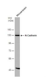
- Western Blot: N-Cadherin Antibody [NBP2-19459] - Mouse tissue extract (50 ug) was separated by 7.5% SDS-PAGE, and the membrane was blotted with N-Cadherin antibody [N1N3] diluted at 1:500. The HRP-conjugated anti-rabbit IgG antibody (NBP2-19301) was used to detect the primary antibody.
- Submitted by
- Novus Biologicals (provider)
- Main image
- Experimental details
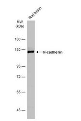
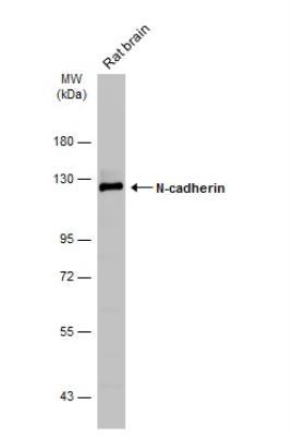
- Western Blot: N-Cadherin Antibody [NBP2-19459] - Rat tissue extract (50 ug) was separated by 7.5% SDS-PAGE, and the membrane was blotted with N-cadherin antibody ( diluted at 1:1000. The HRP-conjugated anti-rabbit IgG antibody (NBP2-19301) was used to detect the primary antibody.
- Submitted by
- Novus Biologicals (provider)
- Main image
- Experimental details
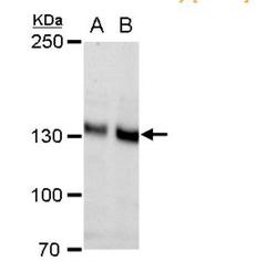
- Western Blot: N-Cadherin Antibody [NBP2-19459] - A. 30 ug PC-12 whole cell extract B. 30 ug Rat2 whole cell extract 5% SDS-PAGE N-Cadherin antibody [N1N3] dilution: 1:1000 The HRP-conjugated anti-rabbit IgG antibody (NBP2-19301) was used to detect the primary antibody.

- Submitted by
- Novus Biologicals (provider)
- Main image
- Experimental details
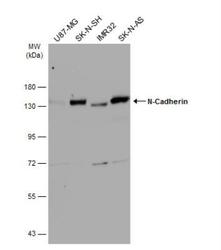
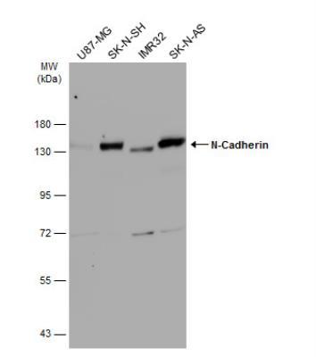
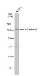
- Western Blot: N-Cadherin Antibody [NBP2-19459] - Various whole cell extracts (30 ug) were separated by 7.5% SDS-PAGE, and the membrane was blotted with N-Cadherin antibody [N1N3] diluted at 1:1000. The HRP-conjugated anti-rabbit IgG antibody (NBP2-19301) was used to detect the primary antibody.
- Submitted by
- Novus Biologicals (provider)
- Main image
- Experimental details
- Western Blot: N-Cadherin Antibody [NBP2-19459] - Whole cell extract (30 ug) was separated by 7.5% SDS-PAGE, and the membrane was blotted with N-Cadherin antibody [N1N3] diluted at 1:1000. The HRP-conjugated anti-rabbit IgG antibody (NBP2-19301) was used to detect the primary antibody.

- Submitted by
- Novus Biologicals (provider)
- Main image
- Experimental details
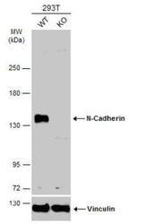
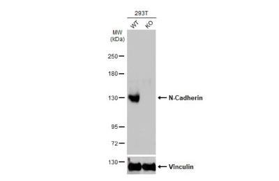
- Western Blot: N-Cadherin Antibody [NBP2-19459] - Wild-type (WT) and N-Cadherin knockout (KO) 293T cell extracts (30 ug) were separated by 5% SDS-PAGE, and the membrane was blotted with N-Cadherin antibody diluted at 1:500. HRP-conjugated anti-rabbit IgG antibody was used to detect the primary antibody.
- Submitted by
- Novus Biologicals (provider)
- Main image
- Experimental details
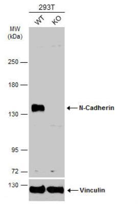
- Western Blot: N-Cadherin Antibody [NBP2-19459] - Wild-type (WT) and N-Cadherin knockout (KO) 293T cell extracts (30 ug) were separated by 5% SDS-PAGE, and the membrane was blotted with N-Cadherin antibody diluted at 1:500. HRP-conjugated anti-rabbit IgG antibody was used to detect the primary antibody.
Supportive validation
- Submitted by
- Novus Biologicals (provider)
- Main image

- Experimental details
- Immunohistochemistry-Paraffin: N Cadherin Antibody [NBP2-19459] - Immunohistochemical analysis of paraffin-embedded Mahlarvu xenograft, using antibody at 1:500 dilution.

- Submitted by
- Novus Biologicals (provider)
- Main image

- Experimental details
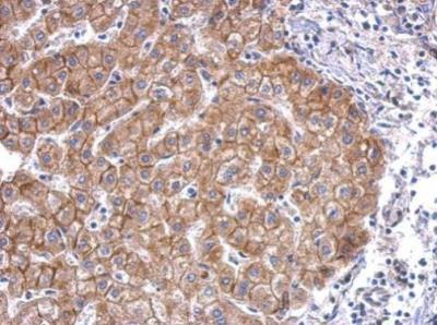
- Immunohistochemistry-Paraffin: N-Cadherin Antibody [NBP2-19459] - Paraffin-embedded human hepatoma. N-Cadherin antibody [N1N3] dilution: 1:500.
- Submitted by
- Novus Biologicals (provider)
- Main image

- Experimental details
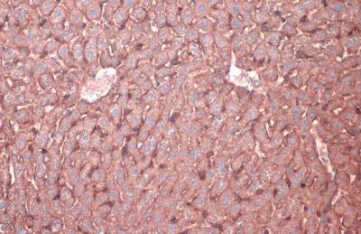
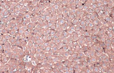
- Immunohistochemistry-Paraffin: N-Cadherin Antibody [NBP2-19459] - N-Cadherin antibody detects N-Cadherin protein at cell membrane by immunohistochemical analysis. Sample: Paraffin-embedded rat liver. N-Cadherin stained by N-Cadherin antibody diluted at 1:500. Antigen Retrieval: Citrate buffer, pH 6.0, 15 min
- Submitted by
- Novus Biologicals (provider)
- Main image

- Experimental details
- Immunohistochemistry-Paraffin: N-Cadherin Antibody [NBP2-19459] - N-Cadherin antibody detects N-Cadherin protein at cell membrane by immunohistochemical analysis. Sample: Paraffin-embedded mouse liver. N-Cadherin stained by N-Cadherin antibody diluted at 1:500. Antigen Retrieval: Citrate buffer, pH 6.0, 15 min.