 Explore
 Validate
 Learn
Learn Western blot
Antibody data
- Antibody Data
- Antigen structure
- References [2]
- Comments [0]
- Validations
- Western blot [1]
- Immunohistochemistry [1]
- Flow cytometry [1]
Submit
Validation data
Reference
Comment
Report error
- Product number
- MAB13881 - Provider product page

- Provider
- Novus Biologicals
- Product name
- Mouse Monoclonal N-Cadherin Antibody
- Antibody type
- Monoclonal
- Description
- Protein A or G purified from hybridoma culture supernatant. Detects mouse N-Cadherin in direct ELISAs and Western blots. In direct ELISAs approximately 50% cross-reactivity with recombinant mouse N-Cadherin is observed, and no cross-reactivity with recombinant human (rh) E-Cadherin, rhP-Cadherin, rhVE-Cadherin, rhCadherin-4, -8, -11, -12, or -13 is observed.
- Reactivity
- Human
- Host
- Mouse
- Conjugate
- Unconjugated
- Isotype
- IgM
- Vial size
- 100 ug
- Concentration
- LYOPH
- Storage
- Use a manual defrost freezer and avoid repeated freeze-thaw cycles. 12 months from date of receipt, -20 to -70 degreesC as supplied. 1 month, 2 to 8 degreesC under sterile conditions after reconstitution. 6 months, -20 to -70 degreesC under sterile conditions after reconstitution.
Submitted references BDNF/TrkB axis activation promotes epithelial-mesenchymal transition in idiopathic pulmonary fibrosis.
Reelin is involved in transforming growth factor-β1-induced cell migration in esophageal carcinoma cells.
Cherubini E, Mariotta S, Scozzi D, Mancini R, Osman G, D'Ascanio M, Bruno P, Cardillo G, Ricci A
Journal of translational medicine 2017 Sep 22;15(1):196
Journal of translational medicine 2017 Sep 22;15(1):196
Reelin is involved in transforming growth factor-β1-induced cell migration in esophageal carcinoma cells.
Yuan Y, Chen H, Ma G, Cao X, Liu Z
PloS one 2012;7(2):e31802
PloS one 2012;7(2):e31802
No comments: Submit comment
Supportive validation
- Submitted by
- Novus Biologicals (provider)
- Main image

- Experimental details
- Detection of Human, Mouse, and Rat N-Cadherin by Western Blot. Western blot shows lysates of A549 human lung carcinoma cell line, C2C12 mouse myoblast cell line, and rat brain tissue. PVDF membrane was probed with 1 µg/mL of Mouse Anti-Human N-Cadherin Monoclonal Antibody (Catalog # MAB13881) followed by HRP-conjugated Anti-Mouse IgG Secondary Antibody (Catalog # HAF007). A specific band was detected for N-Cadherin at approximately 130 kDa (as indicated). This experiment was conducted under reducing conditions and using Immunoblot Buffer Group 1.

Supportive validation
- Submitted by
- Novus Biologicals (provider)
- Main image
- Experimental details
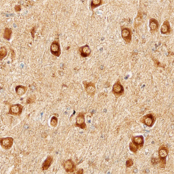
- N-Cadherin in Human Brain. N-Cadherin was detected in immersion fixed paraffin-embedded sections of Alzheimer's human brain using Mouse Anti-Human N-Cadherin Monoclonal Antibody (Catalog # MAB13881) at 15 µg/mL overnight at 4 °C. Before incubation with the primary antibody, tissue was subjected to heat-induced epitope retrieval using Antigen Retrieval Reagent-Basic (Catalog # CTS013). Tissue was stained using the Anti-Mouse HRP-DAB Cell & Tissue Staining Kit (brown; Catalog # CTS002) and counterstained with hematoxylin (blue). Specific staining was localized to cytoplasm of neurons. View our protocol for Chromogenic IHC Staining of Paraffin-embedded Tissue Sections.

Supportive validation
- Submitted by
- Novus Biologicals (provider)
- Main image
- Experimental details
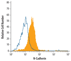
- Detection of N-Cadherin in HeLa Human Cell Line by Flow Cytometry. HeLa human cervical epithelial carcinoma cell line was stained with Mouse Anti-Human N-Cadherin Monoclonal Antibody (Catalog # MAB13881, filled histogram) or isotype control antibody mouse IgM (open histogram), followed by Phycoerythrin-conjugated Anti-Mouse IgM Secondary Antibody (Catalog # F0116).
