 Explore
 Validate
 Learn
Learn Western blot
Antibody data
- Antibody Data
- Antigen structure
- References [0]
- Comments [0]
- Validations
- Western blot [2]
- Immunocytochemistry [1]
- Immunoprecipitation [1]
- Immunohistochemistry [1]
Submit
Validation data
Reference
Comment
Report error
- Product number
- GTX19348 - Provider product page

- Provider
- GeneTex
- Proper citation
- GeneTex Cat#GTX19348, RRID:AB_423537
- Product name
- N-Cadherin antibody [8C11]
- Antibody type
- Monoclonal
- Reactivity
- Human, Mouse, Chicken/Avian, Hamster, Rabbit
- Host
- Mouse
No comments: Submit comment
Supportive validation
- Submitted by
- GeneTex (provider)
- Main image

- Experimental details
- Western blot analysis of N-Cadherin in 25 ug of various lysates. Proteins were transferred to a PVDF membrane and blocked with 5% BSA/TBST for at least 1 hour. The membrane was probed with N-Cadherin antibody [8C11] at a dilution of 1:1000 overnight at 4¢XC on a rocking platform, washed in TBS-0.1%Tween-20, and probed with a HRP-conjugated secondary antibody for 1 hour. Chemiluminescent detection was performed. Note: analysis indicates both reactive and non-reactive species for N-cadherin.

- Submitted by
- GeneTex (provider)
- Main image

- Experimental details
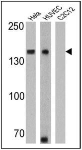
- WB analysis of HeLa, HUVEC and C2C12 lysates (25 ug per lane) using N-Cadherin antibody [8C11] at a dilution of 1:1000 (HeLa) and 1:500 (HUVEC and C2C12).
Supportive validation
- Submitted by
- GeneTex (provider)
- Main image
- Experimental details
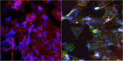
- Immunofluorescent analysis of N-Cadherin in SHSY5Y cells. Formalin-fixed cells were permeabilized with 0.1% Triton X-100 in TBS for 10 minutes at room temperature. Cells were blocked with 1% BSA for 15 minutes at room temperature. Cells were probed without (left panel) or with (right panel) N-Cadherin antibody [8C11] at a dilution of 1:100 for at least 1 hour at room temperature, washed with PBS, and incubated with a proper secondary antibody. F-Actin (red) was stained with Phalloidin and nuclei (blue) were stained with Hoechst 33342 dye. Images were taken at 20X magnification.

Supportive validation
- Submitted by
- GeneTex (provider)
- Main image

- Experimental details
- Immunoprecipitation of N-Cadherin was performed using SHSY5Y whole cell lysate. Antigen-antibody complexes were formed by incubating 300ug of lysate with 5ug of N-Cadherin antibody [8C11] overnight on a rocking platform at 4¢XC. The immune complexes were captured on 50ul Protein A/G Agarose , washed extensively, and eluted. The sample was resolved on a 4-20% Tris-HCl polyacrylamide gel, transferred to a PVDF membrane, and blocked with 5% BSA/TBS-0.1%Tween for at least 1 hour. The membrane was probed with N-Cadherin antibody [8C11] at a dilution of 1:1000 overnight rotating at 4¢XC, washed in TBST, and probed with an proper secondary antibody. Chemiluminescent detection was performed.

Supportive validation
- Submitted by
- GeneTex (provider)
- Main image
- Experimental details
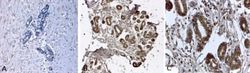
- IHC-P analysis of human breast tissues with an isotype control (A) or N-Cadherin antibody [8C11] (B and C) at a concentration of 2ug/ml. (C) shows the magnified section of (B). To expose target proteins, antigen retrieval was performed using HEIR with a buffer (pH 6.2).
